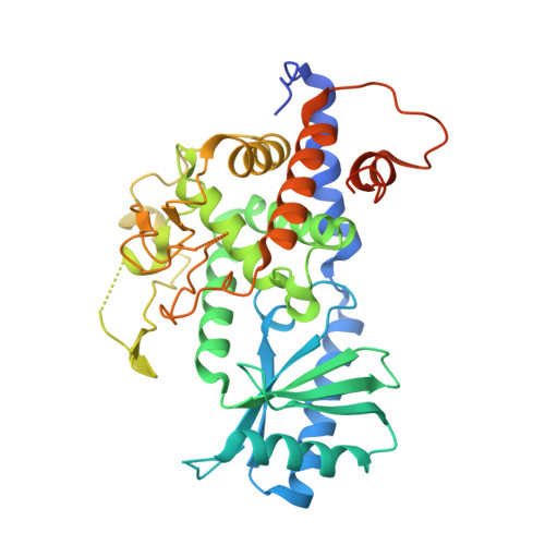
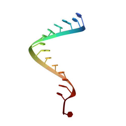
5W0O

Structure of human TUT7 catalytic module (CM) in complex with dsRNA


Experimental Data Snapshot

  • Method: X-RAY DIFFRACTION
  • Resolution: 2.49 Å
  • R-Value Free: 0.254 
  • R-Value Work: 0.205 
  • R-Value Observed: 0.208 

Starting Model: experimental
View more details

wwPDB Validation   3D Report Full Report


Ligand Structure Quality Assessment 


This is version 1.3 of the entry. See complete history


Literature

Multi-domain utilization by TUT4 and TUT7 in control of let-7 biogenesis.

Faehnle, C.R.Walleshauser, J.Joshua-Tor, L.

(2017) Nat Struct Mol Biol 24: 658-665

  • DOI: https://doi.org/10.1038/nsmb.3428
  • Primary Citation of Related Structures:  
    5W0B, 5W0M, 5W0N, 5W0O

  • PubMed Abstract: 

    The uridyl transferases TUT4 and TUT7 (collectively called TUT4(7)) switch between two modes of activity, either promoting expression of let-7 microRNA (monoU) or marking it for degradation (oligoU). Lin28 modulates the switch via recruitment of TUT4(7) to the precursor pre-let-7 in stem cells and human cancers. We found that TUT4(7) utilize two multidomain functional modules during the switch from monoU to oligoU. The catalytic module (CM) is essential for both activities, while the Lin28-interacting module (LIM) is indispensable for oligoU. A TUT7 CM structure trapped in the monoU activity staterevealed a duplex-RNA-binding pocket that orients group II pre-let-7 hairpins to favor monoU addition. Conversely, the switch to oligoU requires the ZK domain of Lin28 to drive the formation of a stable ternary complex between pre-let-7 and the inactive LIM. Finally, ZK2 of TUT4(7) aids oligoU addition by engaging the growing oligoU tail through uracil-specific interactions.


  • Organizational Affiliation

    W.M. Keck Structural Biology Laboratory, Cold Spring Harbor Laboratory, Cold Spring Harbor, New York, USA.


Macromolecules

Find similar proteins by:  (by identity cutoff)  |  3D Structure
Entity ID: 1
MoleculeChains Sequence LengthOrganismDetailsImage
Terminal uridylyltransferase 7
A, B
389Homo sapiensMutation(s): 0 
Gene Names: ZCCHC6HS2KIAA1711TUT7
EC: 2.7.7.52
UniProt & NIH Common Fund Data Resources
Find proteins for Q5VYS8 (Homo sapiens)
Explore Q5VYS8 
Go to UniProtKB:  Q5VYS8
PHAROS:  Q5VYS8
GTEx:  ENSG00000083223 
Entity Groups  
Sequence Clusters30% Identity50% Identity70% Identity90% Identity95% Identity100% Identity
UniProt GroupQ5VYS8
Sequence Annotations
Expand
  • Reference Sequence

Find similar nucleic acids by:  Sequence   |   3D Structure  

Entity ID: 2
MoleculeChains LengthOrganismImage
double-stranded RNA
C, D
15synthetic construct
Sequence Annotations
Expand
  • Reference Sequence
Small Molecules
Ligands 1 Unique
IDChains Name / Formula / InChI Key2D Diagram3D Interactions
UTP
Query on UTP

Download Ideal Coordinates CCD File 
E [auth A],
F [auth B]
URIDINE 5'-TRIPHOSPHATE
C9 H15 N2 O15 P3
PGAVKCOVUIYSFO-XVFCMESISA-N
Experimental Data & Validation

Experimental Data

  • Method: X-RAY DIFFRACTION
  • Resolution: 2.49 Å
  • R-Value Free: 0.254 
  • R-Value Work: 0.205 
  • R-Value Observed: 0.208 
  • Space Group: P 21 21 21
Unit Cell:
Length ( Å )Angle ( ˚ )
a = 66.825α = 90
b = 80.851β = 90
c = 181.83γ = 90
Software Package:
Software NamePurpose
PHENIXrefinement
SCALAdata scaling
PDB_EXTRACTdata extraction
XDSdata reduction
PHASERphasing

Structure Validation

View Full Validation Report


Ligand Structure Quality Assessment 


Entry History 

Deposition Data

Revision History  (Full details and data files)

  • Version 1.0: 2017-06-28
    Type: Initial release
  • Version 1.1: 2017-07-19
    Changes: Database references
  • Version 1.2: 2017-08-16
    Changes: Database references
  • Version 1.3: 2023-10-04
    Changes: Data collection, Database references, Refinement description