5VOO | pdb_00005voo

Methionine synthase folate-binding domain with methyltetrahydrofolate from Thermus thermophilus HB8


Experimental Data Snapshot

  • Method: X-RAY DIFFRACTION
  • Resolution: 2.40 Å
  • R-Value Free: 
    0.230 (Depositor), 0.231 (DCC) 
  • R-Value Work: 
    0.190 (Depositor), 0.192 (DCC) 
  • R-Value Observed: 
    0.192 (Depositor) 

Starting Model: experimental
View more details

wwPDB Validation 3D Report Full Report

Validation slider image for 5VOO

Ligand Structure Quality Assessment 


This is version 1.2 of the entry. See complete history

Literature

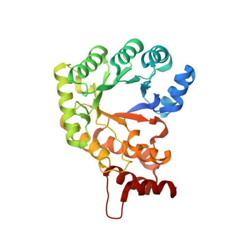
The folate-binding module of Thermus thermophilus cobalamin-dependent methionine synthase displays a distinct variation of the classical TIM barrel: a TIM barrel with a `twist'.

Yamada, K.Koutmos, M.

(2018) Acta Crystallogr D Struct Biol 74: 41-51

  • DOI: https://doi.org/10.1107/S2059798317018290
  • Primary Citation Related Structures: 
    5VON, 5VOO, 5VOP

  • PubMed Abstract: 

    Methyl transfer between methyltetrahydrofolate and corrinoid molecules is a key reaction in biology that is catalyzed by a number of enzymes in many prokaryotic and eukaryotic organisms. One classic example of such an enzyme is cobalamin-dependent methionine synthase (MS). MS is a large modular protein that utilizes an S N 2-type mechanism to catalyze the chemically challenging methyl transfer from the tertiary amine (N5) of methyltetrahydrofolate to homocysteine in order to form methionine. Despite over half a century of study, many questions remain about how folate-dependent methyltransferases, and MS in particular, function. Here, the structure of the folate-binding (Fol) domain of MS from Thermus thermophilus is reported in the presence and absence of methyltetrahydrofolate. It is found that the methyltetrahydrofolate-binding environment is similar to those of previously described methyltransferases, highlighting the conserved role of this domain in binding, and perhaps activating, the methyltetrahydrofolate substrate. These structural studies further reveal a new distinct and uncharacterized topology in the C-terminal region of MS Fol domains. Furthermore, it is found that in contrast to the canonical TIM-barrel β 8 α 8 fold found in all other folate-binding domains, MS Fol domains exhibit a unique β 8 α 7 fold. It is posited that these structural differences are important for MS function.


  • Organizational Affiliation
    • Department of Chemistry, University of Michigan, Ann Arbor, Michigan, USA.

Macromolecule Content 

  • Total Structure Weight: 201.46 kDa 
  • Atom Count: 13,827 
  • Modeled Residue Count: 1,687 
  • Deposited Residue Count: 1,776 
  • Unique protein chains: 1

Macromolecules

Find similar proteins by:|  3D Structure
Entity ID: 1
MoleculeChains  Sequence LengthOrganismDetailsImage
5-methyltetrahydrofolate homocysteine S-methyltransferase
A, B, C, D, E
A, B, C, D, E, F
296Thermus thermophilus HB8Mutation(s): 0 
Gene Names: TTHA0618
EC: 2.1.1.13
UniProt
Find proteins for Q5SKM5 (Thermus thermophilus (strain ATCC 27634 / DSM 579 / HB8))
Explore Q5SKM5 
Go to UniProtKB:  Q5SKM5
Entity Groups
Sequence Clusters30% Identity50% Identity70% Identity90% Identity95% Identity100% Identity
UniProt GroupQ5SKM5
Sequence Annotations
Expand
Reference Sequence

Small Molecules

Ligands 5 Unique
IDChains Name / Formula / InChI Key2D Diagram3D Interactions
C2F

Query on C2F


Download:Ideal Coordinates CCD File
G [auth A]
M [auth B]
Q [auth C]
R [auth D]
U [auth E]
G [auth A],
M [auth B],
Q [auth C],
R [auth D],
U [auth E],
W [auth F]
5-METHYL-5,6,7,8-TETRAHYDROFOLIC ACID
C20 H25 N7 O6
ZNOVTXRBGFNYRX-STQMWFEESA-N
PEG

Query on PEG


Download:Ideal Coordinates CCD File
T [auth D],
V [auth E]
DI(HYDROXYETHYL)ETHER
C4 H10 O3
MTHSVFCYNBDYFN-UHFFFAOYSA-N
GOL

Query on GOL


Download:Ideal Coordinates CCD File
L [auth B],
O [auth B]
GLYCEROL
C3 H8 O3
PEDCQBHIVMGVHV-UHFFFAOYSA-N
CL

Query on CL


Download:Ideal Coordinates CCD File
K [auth A],
P [auth C]
CHLORIDE ION
Cl
VEXZGXHMUGYJMC-UHFFFAOYSA-M
NA

Query on NA


Download:Ideal Coordinates CCD File
H [auth A]
I [auth A]
J [auth A]
N [auth B]
S [auth D]
H [auth A],
I [auth A],
J [auth A],
N [auth B],
S [auth D],
X [auth F]
SODIUM ION
Na
FKNQFGJONOIPTF-UHFFFAOYSA-N

Experimental Data & Validation

Experimental Data

  • Method: X-RAY DIFFRACTION
  • Resolution: 2.40 Å
  • R-Value Free:  0.230 (Depositor), 0.231 (DCC) 
  • R-Value Work:  0.190 (Depositor), 0.192 (DCC) 
  • R-Value Observed: 0.192 (Depositor) 
Space Group: P 61 2 2
Unit Cell:
Length ( Å )Angle ( ˚ )
a = 188.463α = 90
b = 188.463β = 90
c = 247.137γ = 120
Software Package:
Software NamePurpose
REFMACrefinement
Aimlessdata scaling
XDSdata reduction
PHASERphasing

Structure Validation

View Full Validation Report


Ligand Structure Quality Assessment 


Entry History 

& Funding Information

Deposition Data


Funding OrganizationLocationGrant Number
American Heart AssociationUnited States13SDG14560056

Revision History  (Full details and data files)

  • Version 1.0: 2018-01-17
    Type: Initial release
  • Version 1.1: 2018-05-16
    Changes: Data collection, Database references
  • Version 1.2: 2023-10-04
    Changes: Data collection, Database references, Derived calculations, Refinement description