


Bacillus pumilus family 48 glycoside hydrolase in complex with cellobio-derived isofagomine
Alahuhta, P.M., Lunin, V.V.To be published.
Experimental Data Snapshot
Starting Model: experimental
View more details
Entity ID: 1 | |||||
|---|---|---|---|---|---|
| Molecule | Chains | Sequence Length | Organism | Details | Image |
| Glycoside hydrolase | 709 | Bacillus pumilus | Mutation(s): 0 Gene Names: BPUM_1559 | 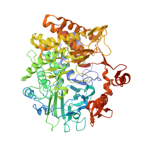 | |
UniProt | |||||
Entity Groups | |||||
| Sequence Clusters | 30% Identity50% Identity70% Identity90% Identity95% Identity100% Identity | ||||
| UniProt Group | A8FDC4 | ||||
Sequence AnnotationsExpand | |||||
Reference Sequence | |||||
| Ligands 7 Unique | |||||
|---|---|---|---|---|---|
| ID | Chains | Name / Formula / InChI Key | 2D Diagram | 3D Interactions | |
| G2I Download:Ideal Coordinates CCD File | B [auth A], C [auth A] | (3R,4R,5R)-3-hydroxy-5-(hydroxymethyl)piperidin-4-yl 4-O-beta-D-glucopyranosyl-beta-D-glucopyranoside C18 H33 N O13 ZIMIYDWQNDRGNF-IONZOCAKSA-N |  | ||
| 9MR Download:Ideal Coordinates CCD File | D [auth A] | (3R,4R,5R)-3-hydroxy-5-(hydroxymethyl)piperidin-4-yl beta-D-glucopyranoside C12 H23 N O8 LEOSSOWHBSKZSO-WUYFHPBOSA-N |  | ||
| MLI Download:Ideal Coordinates CCD File | T [auth A] | MALONATE ION C3 H2 O4 OFOBLEOULBTSOW-UHFFFAOYSA-L |  | ||
| EDO Download:Ideal Coordinates CCD File | H [auth A] I [auth A] J [auth A] K [auth A] L [auth A] | 1,2-ETHANEDIOL C2 H6 O2 LYCAIKOWRPUZTN-UHFFFAOYSA-N |  | ||
| ACT Download:Ideal Coordinates CCD File | U [auth A] | ACETATE ION C2 H3 O2 QTBSBXVTEAMEQO-UHFFFAOYSA-M |  | ||
| CA Download:Ideal Coordinates CCD File | E [auth A], F [auth A] | CALCIUM ION Ca BHPQYMZQTOCNFJ-UHFFFAOYSA-N |  | ||
| NA Download:Ideal Coordinates CCD File | G [auth A] | SODIUM ION Na FKNQFGJONOIPTF-UHFFFAOYSA-N |  | ||
| Length ( Å ) | Angle ( ˚ ) |
|---|---|
| a = 98.891 | α = 90 |
| b = 98.891 | β = 90 |
| c = 216.799 | γ = 90 |
| Software Name | Purpose |
|---|---|
| REFMAC | refinement |
| PROTEUM PLUS | data collection |
| PROTEUM PLUS | data processing |
| MOLREP | phasing |
| Funding Organization | Location | Grant Number |
|---|---|---|
| Department of Energy (DOE, United States) | United States | -- |


