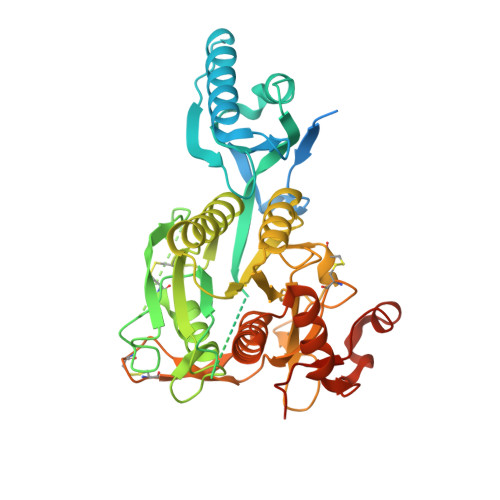
5VLL

Short PCSK9 delta-P' complex with peptide Pep3


Experimental Data Snapshot

  • Method: X-RAY DIFFRACTION
  • Resolution: 2.37 Å
  • R-Value Free: 0.244 
  • R-Value Work: 0.182 
  • R-Value Observed: 0.184 

Starting Model: experimental
View more details

wwPDB Validation   3D Report Full Report


This is version 1.5 of the entry. See complete history


Literature

Discovery of a cryptic peptide-binding site on PCSK9 and design of antagonists.

Zhang, Y.Ultsch, M.Skelton, N.J.Burdick, D.J.Beresini, M.H.Li, W.Kong-Beltran, M.Peterson, A.Quinn, J.Chiu, C.Wu, Y.Shia, S.Moran, P.Di Lello, P.Eigenbrot, C.Kirchhofer, D.

(2017) Nat Struct Mol Biol 24: 848-856

  • DOI: https://doi.org/10.1038/nsmb.3453
  • Primary Citation of Related Structures:  
    5VL7, 5VLA, 5VLH, 5VLK, 5VLL, 5VLP

  • PubMed Abstract: 

    Proprotein convertase subtilisin/kexin type 9 (PCSK9) regulates plasma LDL cholesterol (LDL-c) levels by promoting the degradation of liver LDL receptors (LDLRs). Antibodies that inhibit PCSK9 binding to the EGF(A) domain of the LDLR are effective in lowering LDL-c. However, the discovery of small-molecule therapeutics is hampered by difficulty in targeting the relatively flat EGF(A)-binding site on PCSK9. Here we demonstrate that it is possible to target this site, based on the finding that the PCSK9 P' helix displays conformational flexibility. As a consequence, the vacated N-terminal groove of PCSK9, which is adjacent to the EGF(A)-binding site, is in fact accessible to small peptides. In phage-display experiments, the EGF(A)-mimicking peptide Pep2-8 was used as an anchor peptide for the attachment of an extension peptide library directed toward the groove site. Guided by structural information, we further engineered the identified groove-binding peptides into antagonists, which encroach on the EGF(A)-binding site and inhibit LDLR binding.


  • Organizational Affiliation

    Department of Early Discovery Biochemistry, Genentech, Inc., South San Francisco, California, USA.


Macromolecules
Find similar proteins by:  (by identity cutoff)  |  3D Structure
Entity ID: 1
MoleculeChains Sequence LengthOrganismDetailsImage
Proprotein convertase subtilisin/kexin type 9460Homo sapiensMutation(s): 1 
Gene Names: PCSK9NARC1PSEC0052
EC: 3.4.21
UniProt & NIH Common Fund Data Resources
Find proteins for Q8NBP7 (Homo sapiens)
Go to UniProtKB:  Q8NBP7
PHAROS:  Q8NBP7
GTEx:  ENSG00000169174 
Sequence Annotations
Expand
  • Reference Sequence

Find similar proteins by:  Sequence   |   3D Structure  

Entity ID: 2
MoleculeChains Sequence LengthOrganismDetailsImage
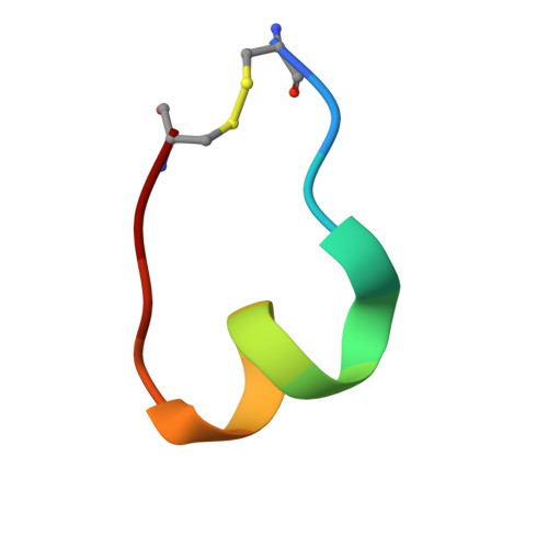
CYS-PHE-ILE-PRO-TRP-ASN-LEU-GLN-ARG-ILE-GLY-LEU-LEU-CYSB [auth Y]14synthetic constructMutation(s): 0 
Sequence Annotations
Expand
  • Reference Sequence

Find similar proteins by:  Sequence   |   3D Structure  

Entity ID: 3
MoleculeChains Sequence LengthOrganismDetailsImage
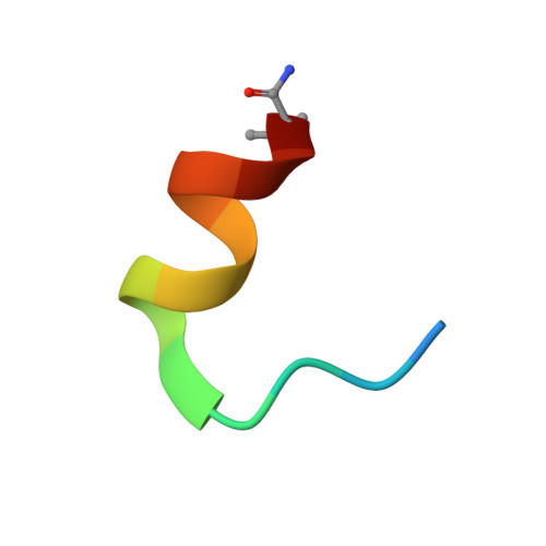
ACE-THR-VAL-PHE-THR-SER-TRP-GLU-GLU-TYR-LEU-ASP-TRP-VAL-NH2C [auth Z]15synthetic constructMutation(s): 0 
Sequence Annotations
Expand
  • Reference Sequence
Small Molecules
Ligands 1 Unique
IDChains Name / Formula / InChI Key2D Diagram3D Interactions
CA
Query on CA

Download Ideal Coordinates CCD File 
D [auth A]CALCIUM ION
Ca
BHPQYMZQTOCNFJ-UHFFFAOYSA-N
Experimental Data & Validation

Experimental Data

  • Method: X-RAY DIFFRACTION
  • Resolution: 2.37 Å
  • R-Value Free: 0.244 
  • R-Value Work: 0.182 
  • R-Value Observed: 0.184 
  • Space Group: P 32 2 1
Unit Cell:
Length ( Å )Angle ( ˚ )
a = 70.566α = 90
b = 70.566β = 90
c = 158.487γ = 120
Software Package:
Software NamePurpose
BUSTERrefinement
XDSdata reduction
Aimlessdata scaling
PHASERphasing

Structure Validation

View Full Validation Report


Entry History & Funding Information

Deposition Data


Funding OrganizationLocationGrant Number
Department of Energy (DOE, United States)United StatesDE-AC02-05CH11231
Department of Energy (DOE, United States)United StatesDE-AC02-06CH11357
National Institutes of Health/National Institute of General Medical Sciences (NIH/NIGMS)United StatesP41GM103393

Revision History  (Full details and data files)

  • Version 1.0: 2017-08-16
    Type: Initial release
  • Version 1.1: 2017-09-06
    Changes: Database references
  • Version 1.2: 2017-09-20
    Changes: Author supporting evidence
  • Version 1.3: 2017-10-18
    Changes: Database references
  • Version 1.4: 2019-12-04
    Changes: Author supporting evidence, Source and taxonomy
  • Version 1.5: 2023-10-04
    Changes: Data collection, Database references, Derived calculations, Refinement description