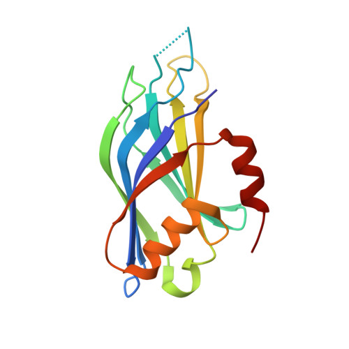
5VFF

Synaptotagmin 1 C2B domain, lead-bound (low occupancy)


Experimental Data Snapshot

  • Method: X-RAY DIFFRACTION
  • Resolution: 1.41 Å
  • R-Value Free: 0.191 
  • R-Value Work: 0.155 
  • R-Value Observed: 0.158 

Starting Model: experimental
View more details

wwPDB Validation   3D Report Full Report


This is version 1.2 of the entry. See complete history


Literature

High affinity interactions of Pb2+with synaptotagmin I.

Katti, S.Her, B.Srivastava, A.K.Taylor, A.B.Lockless, S.W.Igumenova, T.I.

(2018) Metallomics 10: 1211-1222

  • DOI: https://doi.org/10.1039/c8mt00135a
  • Primary Citation of Related Structures:  
    5VFE, 5VFF, 5VFG

  • PubMed Abstract: 

    Lead (Pb) is a potent neurotoxin that disrupts synaptic neurotransmission. We report that Synaptotagmin I (SytI), a key regulator of Ca2+-evoked neurotransmitter release, has two high-affinity Pb2+ binding sites that belong to its cytosolic C2A and C2B domains. The crystal structures of Pb2+-complexed C2 domains revealed that protein-bound Pb2+ ions have holodirected coordination geometries and all-oxygen coordination spheres. The on-rate constants of Pb2+ binding to the C2 domains of SytI are comparable to those of Ca2+ and are diffusion-limited. In contrast, the off-rate constants are at least two orders of magnitude smaller, indicating that Pb2+ can serve as both a thermodynamic and kinetic trap for the C2 domains. We demonstrate, using NMR spectroscopy, that population of these sites by Pb2+ ions inhibits further Ca2+ binding despite the existing coordination vacancies. Our work offers a unique insight into the bioinorganic chemistry of Pb(ii) and suggests a mechanism by which low concentrations of Pb2+ ions can interfere with the Ca2+-dependent function of SytI in the cell.


  • Organizational Affiliation

    Department of Biochemistry and Biophysics, Texas A&M University, 300 Olsen Boulevard, College Station, TX 77843, USA. tigumenova@tamu.edu.


Macromolecules
Find similar proteins by:  (by identity cutoff)  |  3D Structure
Entity ID: 1
MoleculeChains Sequence LengthOrganismDetailsImage
Synaptotagmin-1151Mus musculusMutation(s): 1 
Gene Names: Syt1
UniProt & NIH Common Fund Data Resources
Find proteins for P46096 (Mus musculus)
Explore P46096 
Go to UniProtKB:  P46096
IMPC:  MGI:99667
Entity Groups  
Sequence Clusters30% Identity50% Identity70% Identity90% Identity95% Identity100% Identity
UniProt GroupP46096
Sequence Annotations
Expand
  • Reference Sequence
Small Molecules
Ligands 1 Unique
IDChains Name / Formula / InChI Key2D Diagram3D Interactions
PB
Query on PB

Download Ideal Coordinates CCD File 
B [auth A]LEAD (II) ION
Pb
RVPVRDXYQKGNMQ-UHFFFAOYSA-N
Experimental Data & Validation

Experimental Data

  • Method: X-RAY DIFFRACTION
  • Resolution: 1.41 Å
  • R-Value Free: 0.191 
  • R-Value Work: 0.155 
  • R-Value Observed: 0.158 
  • Space Group: P 21 21 21
Unit Cell:
Length ( Å )Angle ( ˚ )
a = 40.634α = 90
b = 41.117β = 90
c = 82.854γ = 90
Software Package:
Software NamePurpose
PHENIXrefinement
XDSdata reduction
XDSdata scaling
PHASERphasing

Structure Validation

View Full Validation Report


Entry History 

Deposition Data

Revision History  (Full details and data files)

  • Version 1.0: 2018-04-11
    Type: Initial release
  • Version 1.1: 2018-12-19
    Changes: Data collection, Database references
  • Version 1.2: 2023-10-04
    Changes: Data collection, Database references, Derived calculations, Refinement description