5VAJ | pdb_00005vaj

BhRNase H - amide-RNA/DNA complex


Experimental Data Snapshot

  • Method: X-RAY DIFFRACTION
  • Resolution: 1.95 Å
  • R-Value Free: 
    0.217 (Depositor), 0.224 (DCC) 
  • R-Value Work: 
    0.166 (Depositor), 0.175 (DCC) 
  • R-Value Observed: 
    0.168 (Depositor) 

Starting Model: experimental
View more details

wwPDB Validation 3D Report Full Report

Validation slider image for 5VAJ

This is version 1.2 of the entry. See complete history

Literature

Amide linkages mimic phosphates in RNA interactions with proteins and are well tolerated in the guide strand of short interfering RNAs.

Mutisya, D.Hardcastle, T.Cheruiyot, S.K.Pallan, P.S.Kennedy, S.D.Egli, M.Kelley, M.L.Smith, A.V.B.Rozners, E.

(2017) Nucleic Acids Res 45: 8142-8155

  • DOI: https://doi.org/10.1093/nar/gkx558
  • Primary Citation Related Structures: 
    5VAJ

  • PubMed Abstract: 

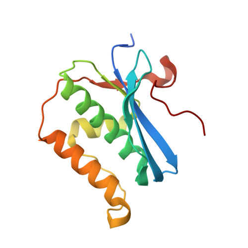
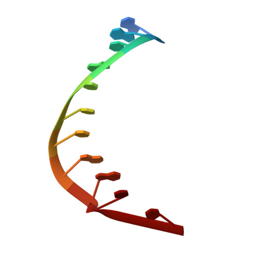
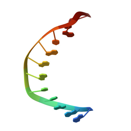
    While the use of RNA interference (RNAi) in molecular biology and functional genomics is a well-established technology, in vivo applications of synthetic short interfering RNAs (siRNAs) require chemical modifications. We recently found that amides as non-ionic replacements for phosphodiesters may be useful modifications for optimization of siRNAs. Herein, we report a comprehensive study of systematic replacement of a single phosphate with an amide linkage throughout the guide strand of siRNAs. The results show that amides are surprisingly well tolerated in the seed and central regions of the guide strand and increase the silencing activity when placed between nucleosides 10 and 12, at the catalytic site of Argonaute. A potential explanation is provided by the first crystal structure of an amide-modified RNA-DNA with Bacillus halodurans RNase H1. The structure reveals how small changes in both RNA and protein conformation allow the amide to establish hydrogen bonding interactions with the protein. Molecular dynamics simulations suggest that these alternative binding modes may compensate for interactions lost due to the absence of a phosphodiester moiety. Our results suggest that an amide can mimic important hydrogen bonding interactions with proteins required for RNAi activity and may be a promising modification for optimization of biological properties of siRNAs.


  • Organizational Affiliation
    • Department of Chemistry, Binghamton University, The State University of New York, Binghamton, NY 13902, USA.

Macromolecule Content 

  • Total Structure Weight: 41.2 kDa 
  • Atom Count: 2,987 
  • Modeled Residue Count: 294 
  • Deposited Residue Count: 307 
  • Unique protein chains: 1
  • Unique nucleic acid chains: 2

Macromolecules


Find similar proteins by:|  3D Structure
Entity ID: 3
MoleculeChains  Sequence LengthOrganismDetailsImage
Ribonuclease HC [auth A],
D [auth B]
142Halalkalibacterium halodurans C-125Mutation(s): 0 
Gene Names: rnhABH0863
EC: 3.1.26.4
UniProt
Find proteins for Q9KEI9 (Halalkalibacterium halodurans (strain ATCC BAA-125 / DSM 18197 / FERM 7344 / JCM 9153 / C-125))
Explore Q9KEI9 
Go to UniProtKB:  Q9KEI9
Entity Groups
Sequence Clusters30% Identity50% Identity70% Identity90% Identity95% Identity100% Identity
UniProt GroupQ9KEI9
Sequence Annotations
Expand
Reference Sequence
Find similar nucleic acids by:  Sequence
Entity ID: 1
MoleculeChains LengthOrganismImage
RNA (5'-R(*GP*AP*CP*AP*CP*CP*UP*GP*AP*(URU)P*C)-3')A [auth C]11synthetic construct
Sequence Annotations
Expand
Reference Sequence
Find similar nucleic acids by:  Sequence
Entity ID: 2
MoleculeChains LengthOrganismImage
DNA (5'-D(*GP*AP*AP*TP*CP*AP*GP*GP*TP*GP*TP*C)-3')B [auth D]12synthetic construct
Sequence Annotations
Expand
Reference Sequence

Small Molecules

Ligands 3 Unique
IDChains Name / Formula / InChI Key2D Diagram3D Interactions
GOL

Query on GOL


Download:Ideal Coordinates CCD File
E [auth C]
F [auth D]
G [auth A]
H [auth A]
I [auth A]
E [auth C],
F [auth D],
G [auth A],
H [auth A],
I [auth A],
J [auth A],
K [auth A],
O [auth B],
P [auth B]
GLYCEROL
C3 H8 O3
PEDCQBHIVMGVHV-UHFFFAOYSA-N
ACT

Query on ACT


Download:Ideal Coordinates CCD File
N [auth A],
S [auth B],
T [auth B]
ACETATE ION
C2 H3 O2
QTBSBXVTEAMEQO-UHFFFAOYSA-M
MG

Query on MG


Download:Ideal Coordinates CCD File
L [auth A],
M [auth A],
Q [auth B],
R [auth B]
MAGNESIUM ION
Mg
JLVVSXFLKOJNIY-UHFFFAOYSA-N

Experimental Data & Validation

Experimental Data

  • Method: X-RAY DIFFRACTION
  • Resolution: 1.95 Å
  • R-Value Free:  0.217 (Depositor), 0.224 (DCC) 
  • R-Value Work:  0.166 (Depositor), 0.175 (DCC) 
  • R-Value Observed: 0.168 (Depositor) 
Space Group: C 1 2 1
Unit Cell:
Length ( Å )Angle ( ˚ )
a = 137.18α = 90
b = 37.513β = 121.65
c = 93.863γ = 90
Software Package:
Software NamePurpose
REFMACrefinement
HKL-2000data reduction
HKL-2000data scaling
MOLREPphasing

Structure Validation

View Full Validation Report


Entry History 

& Funding Information

Deposition Data


Funding OrganizationLocationGrant Number
National Institutes of Health/National Institute of General Medical Sciences (NIH/NIGMS)United StatesR01 GM71461

Revision History  (Full details and data files)

  • Version 1.0: 2017-09-13
    Type: Initial release
  • Version 1.1: 2020-01-01
    Changes: Author supporting evidence
  • Version 1.2: 2023-10-04
    Changes: Data collection, Database references, Derived calculations, Refinement description