5V7T | pdb_00005v7t

crystal structure of PARP14 bound to N-{4-[4-(diphenylmethoxy)piperidin-1-yl]butyl}[1,2,4]triazolo[4,3-b]pyridazin-6-amine inhibitor


Experimental Data Snapshot

  • Method: X-RAY DIFFRACTION
  • Resolution: 2.30 Å
  • R-Value Free: 
    0.291 (Depositor), 0.302 (DCC) 
  • R-Value Work: 
    0.221 (Depositor) 
  • R-Value Observed: 
    0.221 (Depositor) 

wwPDB Validation 3D Report Full Report

Validation slider image for 5V7T

Ligand Structure Quality Assessment 


This is version 1.1 of the entry. See complete history

Literature

Identification of PARP14 inhibitors using novel methods for detecting auto-ribosylation.

Yoneyama-Hirozane, M.Matsumoto, S.I.Toyoda, Y.Saikatendu, K.S.Zama, Y.Yonemori, K.Oonishi, M.Ishii, T.Kawamoto, T.

(2017) Biochem Biophys Res Commun 486: 626-631

  • DOI: https://doi.org/10.1016/j.bbrc.2017.03.052
  • Primary Citation Related Structures: 
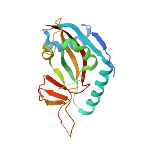
    5V7T, 5V7W

  • PubMed Abstract: 

    Poly(ADP-ribose) polymerases (PARPs) use nicotinamide adenine dinucleotide (NAD + ) as a co-substrate to transfer ADP-ribose when it releases nicotinamide as the metabolized product. Enzymes of the PARP family play key roles in detecting and repairing DNA, modifying chromatin, regulating transcription, controlling energy metabolism, and inducing cell death. PARP14, the original member of the PARP family, has been reported to be associated with the development of inflammatory diseases and various cancer types, making it a potential therapeutic target. In this study, we purified the macrodomain-containing PARP14 enzyme and established an assay for detecting the auto-ribosylation activity of PARP14 using RapidFire high-throughput mass spectrometry and immunoradiometric assay using [ 3 H]NAD + . Subsequently, we performed high-throughput screening using the assays and identified small-molecule hit compounds, which showed NAD + -competitive and PARP14-selective inhibitory activities. Co-crystal structures of PARP14 with certain hit compounds revealed that the inhibitors bind to the NAD + -binding site. Finally, we confirmed that the hit compounds interacted with intracellular PARP14 by a cell-based protein stabilization assay. Thus, we successfully identified primary candidate compounds for further investigation.


  • Organizational Affiliation
    • Pharmaceutical Research Division, Takeda Pharmaceutical Company Limited, 26-1, Muraoka-Higashi 2-chome, Fujisawa, Kanagawa 251-8555, Japan. Electronic address: mariko.hirozane@takeda.com.

Macromolecule Content 

  • Total Structure Weight: 22.56 kDa 
  • Atom Count: 1,507 
  • Modeled Residue Count: 173 
  • Deposited Residue Count: 194 
  • Unique protein chains: 1

Macromolecules

Find similar proteins by:|  3D Structure
Entity ID: 1
MoleculeChains  Sequence LengthOrganismDetailsImage
Poly [ADP-ribose] polymerase 14194Homo sapiensMutation(s): 0 
Gene Names: PARP14BAL2KIAA1268
EC: 2.4.2.30 (PDB Primary Data), 2.4.2 (UniProt)
UniProt & NIH Common Fund Data Resources
Find proteins for Q460N5 (Homo sapiens)
Explore Q460N5 
Go to UniProtKB:  Q460N5
PHAROS:  Q460N5
GTEx:  ENSG00000173193 
Entity Groups
Sequence Clusters30% Identity50% Identity70% Identity90% Identity95% Identity100% Identity
UniProt GroupQ460N5
Sequence Annotations
Expand
Reference Sequence

Small Molecules

Ligands 1 Unique
IDChains Name / Formula / InChI Key2D Diagram3D Interactions
91V

Query on 91V


Download:Ideal Coordinates CCD File
B [auth A]N-{4-[4-(diphenylmethoxy)piperidin-1-yl]butyl}[1,2,4]triazolo[4,3-b]pyridazin-6-amine
C27 H32 N6 O
VPJWDMCKSJWQSU-UHFFFAOYSA-N
Binding Affinity Annotations 
IDSourceBinding Affinity
91V BindingDB:  5V7T IC50: 610 (nM) from 1 assay(s)

Experimental Data & Validation

Experimental Data

  • Method: X-RAY DIFFRACTION
  • Resolution: 2.30 Å
  • R-Value Free:  0.291 (Depositor), 0.302 (DCC) 
  • R-Value Work:  0.221 (Depositor) 
  • R-Value Observed: 0.221 (Depositor) 
Space Group: P 62 2 2
Unit Cell:
Length ( Å )Angle ( ˚ )
a = 83.301α = 90
b = 83.301β = 90
c = 129.94γ = 120
Software Package:
Software NamePurpose
REFMACrefinement
HKL-2000data reduction
HKL-2000data scaling
MOLREPphasing

Structure Validation

View Full Validation Report


Ligand Structure Quality Assessment 


Entry History 

Deposition Data

Revision History  (Full details and data files)

  • Version 1.0: 2017-05-03
    Type: Initial release
  • Version 1.1: 2024-11-06
    Changes: Data collection, Database references, Structure summary