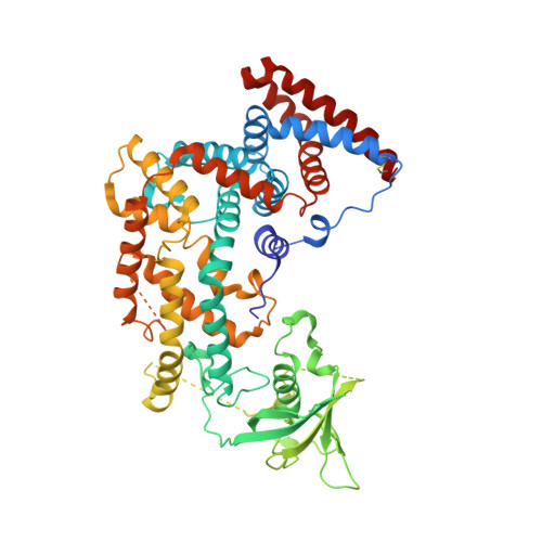
5V6R

Structure of Plexin D1 intracellular domain

  • Classification: PROTEIN BINDING
  • Organism(s): Mus musculus
  • Expression System: Escherichia coli
  • Mutation(s): No 

  • Deposited: 2017-03-17 Released: 2017-05-31 
  • Deposition Author(s): Shang, G., Zhang, X.
  • Funding Organization(s): National Institutes of Health/National Institute of General Medical Sciences (NIH/NIGMS), Welch Foundation

Experimental Data Snapshot

  • Method: X-RAY DIFFRACTION
  • Resolution: 2.70 Å
  • R-Value Free: 0.256 
  • R-Value Work: 0.215 
  • R-Value Observed: 0.216 

Starting Model: experimental
View more details

wwPDB Validation   3D Report Full Report


This is version 1.5 of the entry. See complete history


Literature

Structure analyses reveal a regulated oligomerization mechanism of the PlexinD1/GIPC/myosin VI complex.

Shang, G.Brautigam, C.A.Chen, R.Lu, D.Torres-Vazquez, J.Zhang, X.

(2017) Elife 6

  • DOI: https://doi.org/10.7554/eLife.27322
  • Primary Citation of Related Structures:  
    5V6B, 5V6E, 5V6H, 5V6R, 5V6T

  • PubMed Abstract: 

    The GIPC family adaptor proteins mediate endocytosis by tethering cargo proteins to the myosin VI motor. The structural mechanisms for the GIPC/cargo and GIPC/myosin VI interactions remained unclear. PlexinD1, a transmembrane receptor that regulates neuronal and cardiovascular development, is a cargo of GIPCs. GIPC-mediated endocytic trafficking regulates PlexinD1 signaling. Here, we unravel the mechanisms of the interactions among PlexinD1, GIPCs and myosin VI by a series of crystal structures of these proteins in apo or bound states. GIPC1 forms a domain-swapped dimer in an autoinhibited conformation that hinders binding of both PlexinD1 and myosin VI. PlexinD1 binding to GIPC1 releases the autoinhibition, promoting its interaction with myosin VI. GIPCs and myosin VI interact through two distinct interfaces and form an open-ended alternating array. Our data support that this alternating array underlies the oligomerization of the GIPC/Myosin VI complexes in solution and cells.


  • Organizational Affiliation

    Department of Pharmacology, University of Texas Southwestern Medical Center, Dallas, United States.


Macromolecules
Find similar proteins by:  (by identity cutoff)  |  3D Structure
Entity ID: 1
MoleculeChains Sequence LengthOrganismDetailsImage
Plexin-D1
A, B
587Mus musculusMutation(s): 0 
Gene Names: Plxnd1
UniProt
Find proteins for Q3UH93 (Mus musculus)
Explore Q3UH93 
Go to UniProtKB:  Q3UH93
Entity Groups  
Sequence Clusters30% Identity50% Identity70% Identity90% Identity95% Identity100% Identity
UniProt GroupQ3UH93
Sequence Annotations
Expand
  • Reference Sequence
Experimental Data & Validation

Experimental Data

  • Method: X-RAY DIFFRACTION
  • Resolution: 2.70 Å
  • R-Value Free: 0.256 
  • R-Value Work: 0.215 
  • R-Value Observed: 0.216 
  • Space Group: P 1 21 1
Unit Cell:
Length ( Å )Angle ( ˚ )
a = 69.852α = 90
b = 164.544β = 99.47
c = 84.268γ = 90
Software Package:
Software NamePurpose
PHENIXrefinement
HKL-2000data reduction
HKL-2000data scaling
PHASERphasing

Structure Validation

View Full Validation Report


Entry History & Funding Information

Deposition Data


Funding OrganizationLocationGrant Number
National Institutes of Health/National Institute of General Medical Sciences (NIH/NIGMS)United StatesGM088197
Welch FoundationUnited StatesI-1702

Revision History  (Full details and data files)

  • Version 1.0: 2017-05-31
    Type: Initial release
  • Version 1.1: 2017-06-07
    Changes: Database references
  • Version 1.2: 2017-09-27
    Changes: Author supporting evidence
  • Version 1.3: 2020-01-01
    Changes: Author supporting evidence
  • Version 1.4: 2023-10-04
    Changes: Data collection, Database references, Refinement description
  • Version 1.5: 2024-10-09
    Changes: Structure summary