5V5V

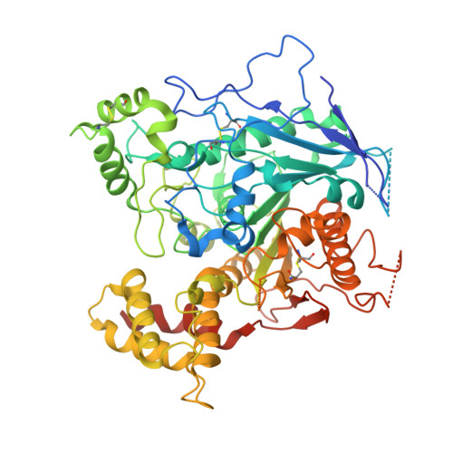
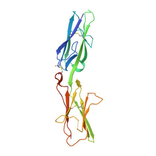
Complex of NLGN2 with MDGA1 Ig1-Ig2

  • Classification: CELL ADHESION
  • Mutation(s): No 

  • Deposited: 2017-03-15 Released: 2017-07-05 
  • Deposition Author(s): Gangwar, S.P., Machius, M., Rudenko, G.
  • Funding Organization(s): National Institutes of Health/National Institute of Mental Health (NIH/NIMH), UT Brain Pilot, Brain and Behavior Research Foundation

Experimental Data Snapshot

  • Method: X-RAY DIFFRACTION
  • Resolution: 4.11 Å
  • R-Value Free: 0.319 
  • R-Value Work: 0.289 
  • R-Value Observed: 0.290 

Starting Model: experimental
View more details

wwPDB Validation   3D Report Full Report


This is version 1.3 of the entry. See complete history


Literature

Molecular Mechanism of MDGA1: Regulation of Neuroligin 2:Neurexin Trans-synaptic Bridges.

Gangwar, S.P.Zhong, X.Seshadrinathan, S.Chen, H.Machius, M.Rudenko, G.

(2017) Neuron 94: 1132-1141.e4

  • DOI: https://doi.org/10.1016/j.neuron.2017.06.009
  • Primary Citation of Related Structures:  
    5V5V, 5V5W

  • PubMed Abstract: 

    Neuroligins and neurexins promote synapse development and validation by forming trans-synaptic bridges spanning the synaptic cleft. Select pairs promote excitatory and inhibitory synapses, with neuroligin 2 (NLGN2) limited to inhibitory synapses and neuroligin 1 (NLGN1) dominating at excitatory synapses. The cell-surface molecules, MAM domain-containing glycosylphosphatidylinositol anchor 1 (MDGA1) and 2 (MDGA2), regulate trans-synaptic adhesion between neurexins and neuroligins, impacting NLGN2 and NLGN1, respectively. We have determined the molecular mechanism of MDGA action. MDGA1 Ig1-Ig2 is sufficient to bind NLGN2 with nanomolar affinity; its crystal structure reveals an unusual locked rod-shaped array. In the crystal structure of the complex, two MDGA1 Ig1-Ig2 molecules each span the entire NLGN2 dimer. Site-directed mutagenesis confirms the observed interaction interface. Strikingly, Ig1 from MDGA1 binds to the same region on NLGN2 as neurexins do. Thus, MDGAs regulate the formation of neuroligin-neurexin trans-synaptic bridges by sterically blocking access of neurexins to neuroligins.


  • Organizational Affiliation

    Department of Pharmacology and Toxicology, University of Texas Medical Branch, Galveston, TX 77555, USA; Sealy Center for Structural Biology and Molecular Biophysics, University of Texas Medical Branch, Galveston, TX 77555, USA.


Macromolecules
Experimental Data & Validation

Experimental Data

  • Method: X-RAY DIFFRACTION
  • Resolution: 4.11 Å
  • R-Value Free: 0.319 
  • R-Value Work: 0.289 
  • R-Value Observed: 0.290 
  • Space Group: P 1
Unit Cell:
Length ( Å )Angle ( ˚ )
a = 103.118α = 95.52
b = 97.182β = 80.97
c = 190.51γ = 88.71
Software Package:
Software NamePurpose
REFMACrefinement
HKL-2000data reduction
HKL-2000data scaling
PHASERphasing

Structure Validation

View Full Validation Report


Entry History & Funding Information

Deposition Data


Funding OrganizationLocationGrant Number
National Institutes of Health/National Institute of Mental Health (NIH/NIMH)United StatesR01MH077303
UT Brain PilotUnited States--
Brain and Behavior Research FoundationUnited States--

Revision History  (Full details and data files)

  • Version 1.0: 2017-07-05
    Type: Initial release
  • Version 1.1: 2017-09-27
    Changes: Author supporting evidence
  • Version 1.2: 2019-11-27
    Changes: Author supporting evidence
  • Version 1.3: 2023-10-04
    Changes: Data collection, Database references, Refinement description