5UWM | pdb_00005uwm

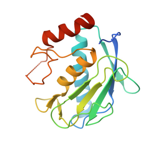
Matrix metalloproteinase-13 complexed with selective inhibitor compound (R)-17a


Experimental Data Snapshot

  • Method: X-RAY DIFFRACTION
  • Resolution: 1.62 Å
  • R-Value Free: 
    0.178 (Depositor), 0.180 (DCC) 
  • R-Value Work: 
    0.129 (Depositor), 0.130 (DCC) 
  • R-Value Observed: 
    0.131 (Depositor) 

Starting Model: experimental
View more details

wwPDB Validation   3D Report Full Report


Ligand Structure Quality Assessment 

Created with Raphaël 2.3.0Worse 01 BetterLigand structure goodness of fit to experimental dataBest fitted 8OAClick on this verticalbar to view details

This is version 1.2 of the entry. See complete history


Literature

Structure-Based Design and Synthesis of Potent and Selective Matrix Metalloproteinase 13 Inhibitors.

Choi, J.Y.Fuerst, R.Knapinska, A.M.Taylor, A.B.Smith, L.Cao, X.Hart, P.J.Fields, G.B.Roush, W.R.

(2017) J Med Chem 60: 5816-5825

  • DOI: https://doi.org/10.1021/acs.jmedchem.7b00514
  • Primary Citation of Related Structures:  
    5UWK, 5UWL, 5UWM, 5UWN

  • PubMed Abstract: 

    We describe the use of comparative structural analysis and structure-guided molecular design to develop potent and selective inhibitors (10d and (S)-17b) of matrix metalloproteinase 13 (MMP-13). We applied a three-step process, starting with a comparative analysis of the X-ray crystallographic structure of compound 5 in complex with MMP-13 with published structures of known MMP-13·inhibitor complexes followed by molecular design and synthesis of potent but nonselective zinc-chelating MMP inhibitors (e.g., 10a and 10b). After demonstrating that the pharmacophores of the chelating inhibitors (S)-10a, (R)-10a, and 10b were binding within the MMP-13 active site, the Zn 2+ chelating unit was replaced with nonchelating polar residues that bridged over the Zn 2+ binding site and reached into a solvent accessible area. After two rounds of structural optimization, these design approaches led to small molecule MMP-13 inhibitors 10d and (S)-17b, which bind within the substrate-binding site of MMP-13 and surround the catalytically active Zn 2+ ion without chelating to the metal. These compounds exhibit at least 500-fold selectivity versus other MMPs.


  • Organizational Affiliation

    Department of Chemistry, Scripps Florida , 130 Scripps Way, Jupiter, Florida 33458, United States.


Macromolecules
Find similar proteins by:  (by identity cutoff)  |  3D Structure
Entity ID: 1
MoleculeChains Sequence LengthOrganismDetailsImage
Collagenase 3
A, B
172Homo sapiensMutation(s): 0 
Gene Names: MMP13
EC: 3.4.24
UniProt & NIH Common Fund Data Resources
Find proteins for P45452 (Homo sapiens)
Explore P45452 
Go to UniProtKB:  P45452
PHAROS:  P45452
GTEx:  ENSG00000137745 
Entity Groups  
Sequence Clusters30% Identity50% Identity70% Identity90% Identity95% Identity100% Identity
UniProt GroupP45452
Sequence Annotations
Expand
  • Reference Sequence

Find similar proteins by:  Sequence   |   3D Structure  

Entity ID: 2
MoleculeChains Sequence LengthOrganismDetailsImage
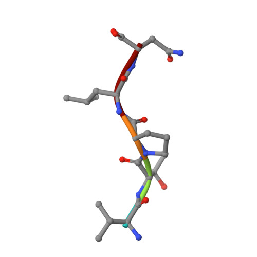
Collagenase 3
C, D
5Homo sapiensMutation(s): 0 
EC: 3.4.24
UniProt & NIH Common Fund Data Resources
Find proteins for P45452 (Homo sapiens)
Explore P45452 
Go to UniProtKB:  P45452
PHAROS:  P45452
GTEx:  ENSG00000137745 
Entity Groups  
UniProt GroupP45452
Sequence Annotations
Expand
  • Reference Sequence
Small Molecules
Ligands 3 Unique
IDChains Name / Formula / InChI Key2D Diagram3D Interactions
8OA
Query on 8OA

Download Ideal Coordinates CCD File 
J [auth A],
P [auth B]
(R)-N-(3-methyl-1-(methylamino)-1-oxobutan-2-yl)-5-(4-(((4-oxo-4,5,6,7-tetrahydro-3H-cyclopenta[d]pyrimidin-2-yl)thio)methyl)phenyl)furan-2-carboxamide
C25 H28 N4 O4 S
AHTVZGHNJWEFAC-OAQYLSRUSA-N
ZN
Query on ZN

Download Ideal Coordinates CCD File 
E [auth A],
F [auth A],
K [auth B],
L [auth B]
ZINC ION
Zn
PTFCDOFLOPIGGS-UHFFFAOYSA-N
CA
Query on CA

Download Ideal Coordinates CCD File 
G [auth A]
H [auth A]
I [auth A]
M [auth B]
N [auth B]
CALCIUM ION
Ca
BHPQYMZQTOCNFJ-UHFFFAOYSA-N
Binding Affinity Annotations 
IDSourceBinding Affinity
8OA BindingDB:  5UWM IC50: 356 (nM) from 1 assay(s)
Experimental Data & Validation

Experimental Data

  • Method: X-RAY DIFFRACTION
  • Resolution: 1.62 Å
  • R-Value Free:  0.178 (Depositor), 0.180 (DCC) 
  • R-Value Work:  0.129 (Depositor), 0.130 (DCC) 
  • R-Value Observed: 0.131 (Depositor) 
Space Group: H 3
Unit Cell:
Length ( Å )Angle ( ˚ )
a = 140.073α = 90
b = 140.073β = 90
c = 46.86γ = 120
Software Package:
Software NamePurpose
PHENIXrefinement
XDSdata reduction
XDSdata scaling
PHASERphasing

Structure Validation

View Full Validation Report


Ligand Structure Quality Assessment 

Created with Raphaël 2.3.0Worse 01 BetterLigand structure goodness of fit to experimental dataBest fitted 8OAClick on this verticalbar to view details

Entry History 

Deposition Data

Revision History  (Full details and data files)

  • Version 1.0: 2017-07-12
    Type: Initial release
  • Version 1.1: 2017-07-26
    Changes: Database references
  • Version 1.2: 2023-10-04
    Changes: Data collection, Database references, Derived calculations, Refinement description