5UV5 | pdb_00005uv5

Crystal Structure of a 2-Hydroxyisoquinoline-1,3-dione RNase H Active Site Inhibitor with Multiple Binding Modes to HIV Reverse Transcriptase


Experimental Data Snapshot

  • Method: X-RAY DIFFRACTION
  • Resolution: 3.00 Å
  • R-Value Free: 
    0.287 (Depositor), 0.296 (DCC) 
  • R-Value Work: 
    0.232 (Depositor), 0.238 (DCC) 
  • R-Value Observed: 
    0.235 (Depositor) 

Starting Model: experimental
View more details

wwPDB Validation 3D Report Full Report

Validation slider image for 5UV5

Ligand Structure Quality Assessment 


This is version 1.4 of the entry. See complete history. 

Literature

A 2-Hydroxyisoquinoline-1,3-Dione Active-Site RNase H Inhibitor Binds in Multiple Modes to HIV-1 Reverse Transcriptase.

Kirby, K.A., Myshakina, N.A., Christen, M.T., Chen, Y.L., Schmidt, H.A., Huber, A.D., Xi, Z., Kim, S., Rao, R.K., Kramer, S.T., Yang, Q., Singh, K., Parniak, M.A., Wang, Z., Ishima, R., Sarafianos, S.G.

(2017) Antimicrob Agents Chemother 61

  • DOI: https://doi.org/10.1128/AAC.01351-17
  • Primary Citation Related Structures: 
    5UV5

  • PubMed Abstract: 

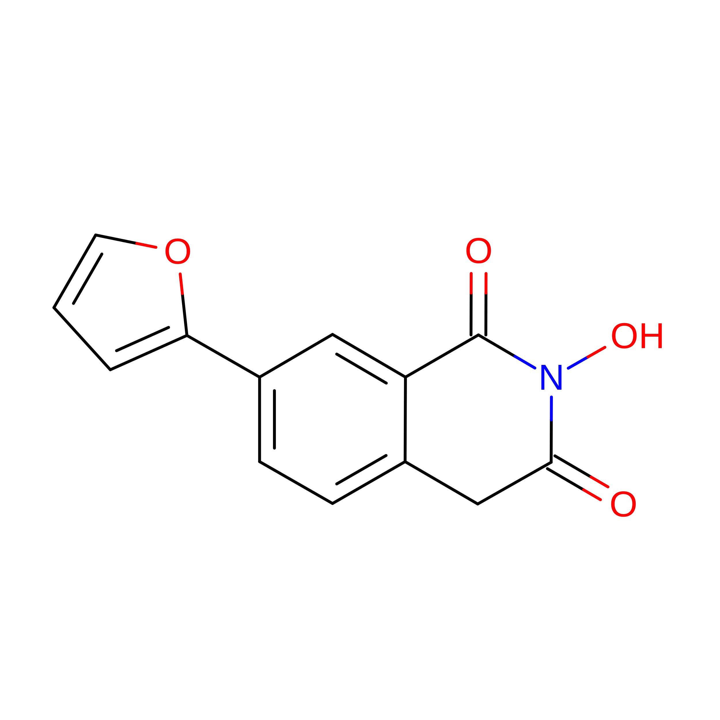
    The RNase H (RNH) function of HIV-1 reverse transcriptase (RT) plays an essential part in the viral life cycle. We report the characterization of YLC2-155, a 2-hydroxyisoquinoline-1,3-dione (HID)-based active-site RNH inhibitor. YLC2-155 inhibits both polymerase (50% inhibitory concentration [IC 50 ] = 2.6 μM) and RNH functions (IC 50 = 0.65 μM) of RT but is more effective against RNH. X-ray crystallography, nuclear magnetic resonance (NMR) analysis, and molecular modeling were used to show that YLC2-155 binds at the RNH-active site in multiple conformations.


  • Organizational Affiliation: 
    • Christopher S. Bond Life Sciences Center, University of Missouri, Columbia, Missouri, USA kirbyk@missouri.edu sarafianoss@missouri.edu.

Macromolecule Content 

  • Total Structure Weight: 229.11 kDa 
  • Atom Count: 15,370 
  • Modeled Residue Count: 1,873 
  • Deposited Residue Count: 1,972 
  • Unique protein chains: 2

Macromolecules

Find similar proteins by:|  3D Structure
Entity ID: 1
MoleculeChains  Sequence LengthOrganismDetailsImage
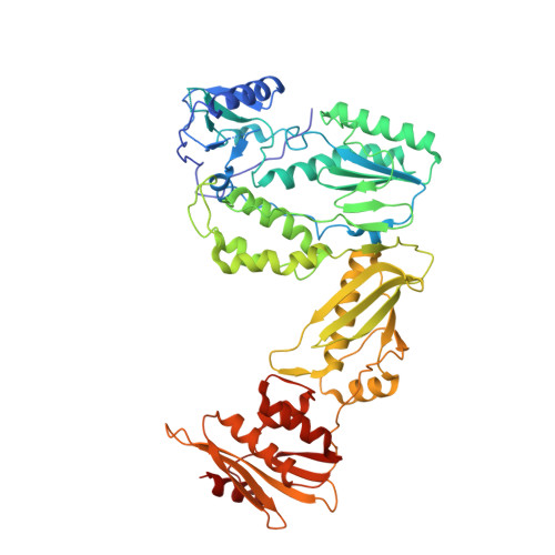
Reverse transcriptase/ribonuclease H
A, C
557Human immunodeficiency virus type 1 BH10Mutation(s): 1 
Gene Names: gag-pol
EC: 2.7.7.49 (PDB Primary Data), 2.7.7.7 (PDB Primary Data), 3.1.26.13 (PDB Primary Data)
UniProt
Find proteins for P03366 (Human immunodeficiency virus type 1 group M subtype B (isolate BH10))
Explore P03366 
Go to UniProtKB:  P03366
Entity Groups
Sequence Clusters30% Identity50% Identity70% Identity90% Identity95% Identity100% Identity
UniProt GroupP03366
Sequence Annotations
Expand
Reference Sequence
Find similar proteins by:|  3D Structure
Entity ID: 2
MoleculeChains  Sequence LengthOrganismDetailsImage
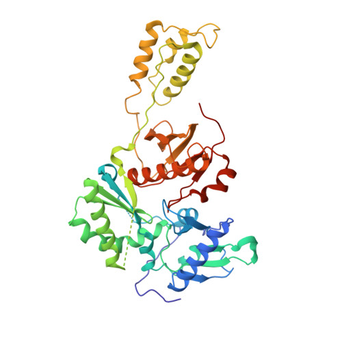
p51 RT
B, D
429Human immunodeficiency virus type 1 BH10Mutation(s): 1 
Gene Names: gag-pol
UniProt
Find proteins for P03366 (Human immunodeficiency virus type 1 group M subtype B (isolate BH10))
Explore P03366 
Go to UniProtKB:  P03366
Entity Groups
Sequence Clusters30% Identity50% Identity70% Identity90% Identity95% Identity100% Identity
UniProt GroupP03366
Sequence Annotations
Expand
Reference Sequence

Experimental Data & Validation

Experimental Data

  • Method: X-RAY DIFFRACTION
  • Resolution: 3.00 Å
  • R-Value Free:  0.287 (Depositor), 0.296 (DCC) 
  • R-Value Work:  0.232 (Depositor), 0.238 (DCC) 
  • R-Value Observed: 0.235 (Depositor) 
Space Group: P 1
Unit Cell:
Length ( Å )Angle ( ˚ )
a = 68.99α = 105.53
b = 88.67β = 93.41
c = 108.86γ = 110.13
Software Package:
Software NamePurpose
Aimlessdata scaling
PHASERphasing
PHENIXrefinement
PDB_EXTRACTdata extraction
XDSdata reduction

Structure Validation

View Full Validation Report


Ligand Structure Quality Assessment 


Entry History 

& Funding Information

Deposition Data


Funding OrganizationLocationGrant Number
National Institutes of Health/National Institute Of Allergy and Infectious Diseases (NIH/NIAID)United StatesAI100890

Revision History  (Full details and data files)

  • Version 1.0: 2017-08-16
    Type: Initial release
  • Version 1.1: 2017-09-20
    Changes: Author supporting evidence
  • Version 1.2: 2017-10-04
    Changes: Database references
  • Version 1.3: 2019-12-11
    Changes: Author supporting evidence
  • Version 1.4: 2023-10-04
    Changes: Data collection, Database references, Derived calculations, Refinement description