5UR5 | pdb_00005ur5

PYR1 bound to the rationally designed agonist 4m


Experimental Data Snapshot

  • Method: X-RAY DIFFRACTION
  • Resolution: 1.93 Å
  • R-Value Free: 
    0.186 (Depositor), 0.187 (DCC) 
  • R-Value Work: 
    0.153 (Depositor), 0.155 (DCC) 
  • R-Value Observed: 
    0.157 (Depositor) 

Starting Model: experimental
View more details

wwPDB Validation 3D Report Full Report

Validation slider image for 5UR5

Ligand Structure Quality Assessment 


This is version 1.2 of the entry. See complete history

Literature

A Rationally Designed Agonist Defines Subfamily IIIA Abscisic Acid Receptors As Critical Targets for Manipulating Transpiration.

Vaidya, A.S.Peterson, F.C.Yarmolinsky, D.Merilo, E.Verstraeten, I.Park, S.Y.Elzinga, D.Kaundal, A.Helander, J.Lozano-Juste, J.Otani, M.Wu, K.Jensen, D.R.Kollist, H.Volkman, B.F.Cutler, S.R.

(2017) ACS Chem Biol 12: 2842-2848

  • DOI: https://doi.org/10.1021/acschembio.7b00650
  • Primary Citation Related Structures: 
    5UR5, 5UR6

  • PubMed Abstract: 

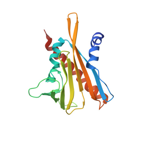
    Increasing drought and diminishing freshwater supplies have stimulated interest in developing small molecules that can be used to control transpiration. Receptors for the plant hormone abscisic acid (ABA) have emerged as key targets for this application, because ABA controls the apertures of stomata, which in turn regulate transpiration. Here, we describe the rational design of cyanabactin, an ABA receptor agonist that preferentially activates Pyrabactin Resistance 1 (PYR1) with low nanomolar potency. A 1.63 Å X-ray crystallographic structure of cyanabactin in complex with PYR1 illustrates that cyanabactin's arylnitrile mimics ABA's cyclohexenone oxygen and engages the tryptophan lock, a key component required to stabilize activated receptors. Further, its sulfonamide and 4-methylbenzyl substructures mimic ABA's carboxylate and C6 methyl groups, respectively. Isothermal titration calorimetry measurements show that cyanabactin's compact structure provides ready access to high ligand efficiency on a relatively simple scaffold. Cyanabactin treatments reduce Arabidopsis whole-plant stomatal conductance and activate multiple ABA responses, demonstrating that its in vitro potency translates to ABA-like activity in vivo. Genetic analyses show that the effects of cyanabactin, and the previously identified agonist quinabactin, can be abolished by the genetic removal of PYR1 and PYL1, which form subclade A within the dimeric subfamily III receptors. Thus, cyanabactin is a potent and selective agonist with a wide spectrum of ABA-like activities that defines subfamily IIIA receptors as key target sites for manipulating transpiration.


  • Organizational Affiliation
    • Department of Botany and Plant Sciences, Institute of Integrative Genome Biology, University of California , Riverside, California 92521, United States.

Macromolecule Content 

  • Total Structure Weight: 22 kDa 
  • Atom Count: 1,629 
  • Modeled Residue Count: 174 
  • Deposited Residue Count: 181 
  • Unique protein chains: 1

Macromolecules

Find similar proteins by:|  3D Structure
Entity ID: 1
MoleculeChains  Sequence LengthOrganismDetailsImage
Abscisic acid receptor PYR1181Arabidopsis thalianaMutation(s): 0 
Gene Names: PYR1ABIP6RCAR11At4g17870T6K21.50
UniProt
Find proteins for O49686 (Arabidopsis thaliana)
Explore O49686 
Go to UniProtKB:  O49686
Entity Groups
Sequence Clusters30% Identity50% Identity70% Identity90% Identity95% Identity100% Identity
UniProt GroupO49686
Sequence Annotations
Expand
Reference Sequence

Small Molecules

Ligands 3 Unique
IDChains Name / Formula / InChI Key2D Diagram3D Interactions
8KP

Query on 8KP


Download:Ideal Coordinates CCD File
H [auth A]N-(4-cyano-3-ethyl-5-methylphenyl)-1-(4-methylphenyl)methanesulfonamide
C18 H20 N2 O2 S
JJJGFSNVNAZOCI-UHFFFAOYSA-N
SO4

Query on SO4


Download:Ideal Coordinates CCD File
B [auth A]
C [auth A]
D [auth A]
E [auth A]
F [auth A]
B [auth A],
C [auth A],
D [auth A],
E [auth A],
F [auth A],
G [auth A]
SULFATE ION
O4 S
QAOWNCQODCNURD-UHFFFAOYSA-L
GOL

Query on GOL


Download:Ideal Coordinates CCD File
I [auth A],
J [auth A],
K [auth A],
L [auth A]
GLYCEROL
C3 H8 O3
PEDCQBHIVMGVHV-UHFFFAOYSA-N

Experimental Data & Validation

Experimental Data

  • Method: X-RAY DIFFRACTION
  • Resolution: 1.93 Å
  • R-Value Free:  0.186 (Depositor), 0.187 (DCC) 
  • R-Value Work:  0.153 (Depositor), 0.155 (DCC) 
  • R-Value Observed: 0.157 (Depositor) 
Space Group: F 2 2 2
Unit Cell:
Length ( Å )Angle ( ˚ )
a = 82.605α = 90
b = 88.22β = 90
c = 112.167γ = 90
Software Package:
Software NamePurpose
PHENIXrefinement
HKL-2000data collection
HKL-2000data scaling
PHASERphasing
PDB_EXTRACTdata extraction
HKL-2000data reduction
HKLdata reduction
HKLdata scaling

Structure Validation

View Full Validation Report


Ligand Structure Quality Assessment 


Entry History 

& Funding Information

Deposition Data


Funding OrganizationLocationGrant Number
National Science Foundation (NSF, United States)United States1258175

Revision History  (Full details and data files)

  • Version 1.0: 2017-11-29
    Type: Initial release
  • Version 1.1: 2019-11-27
    Changes: Author supporting evidence, Derived calculations
  • Version 1.2: 2023-10-04
    Changes: Data collection, Database references, Refinement description