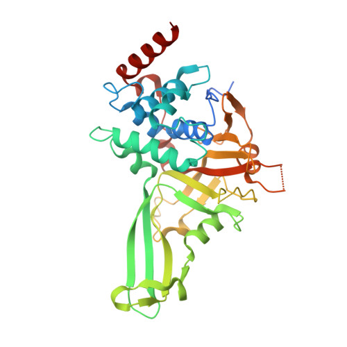
USP7 small-molecule inhibitors interfere with ubiquitin binding.
Kategaya, L., Di Lello, P., Rouge, L., Pastor, R., Clark, K.R., Drummond, J., Kleinheinz, T., Lin, E., Upton, J.P., Prakash, S., Heideker, J., McCleland, M., Ritorto, M.S., Alessi, D.R., Trost, M., Bainbridge, T.W., Kwok, M.C.M., Ma, T.P., Stiffler, Z., Brasher, B., Tang, Y., Jaishankar, P., Hearn, B.R., Renslo, A.R., Arkin, M.R., Cohen, F., Yu, K., Peale, F., Gnad, F., Chang, M.T., Klijn, C., Blackwood, E., Martin, S.E., Forrest, W.F., Ernst, J.A., Ndubaku, C., Wang, X., Beresini, M.H., Tsui, V., Schwerdtfeger, C., Blake, R.A., Murray, J., Maurer, T., Wertz, I.E.(2017) Nature 550: 534-538
- PubMed: 29045385
- DOI: https://doi.org/10.1038/nature24006
- Primary Citation of Related Structures:
5UQV, 5UQX - PubMed Abstract:
The ubiquitin system regulates essential cellular processes in eukaryotes. Ubiquitin is ligated to substrate proteins as monomers or chains and the topology of ubiquitin modifications regulates substrate interactions with specific proteins. Thus ubiquitination directs a variety of substrate fates including proteasomal degradation. Deubiquitinase enzymes cleave ubiquitin from substrates and are implicated in disease; for example, ubiquitin-specific protease-7 (USP7) regulates stability of the p53 tumour suppressor and other proteins critical for tumour cell survival. However, developing selective deubiquitinase inhibitors has been challenging and no co-crystal structures have been solved with small-molecule inhibitors. Here, using nuclear magnetic resonance-based screening and structure-based design, we describe the development of selective USP7 inhibitors GNE-6640 and GNE-6776. These compounds induce tumour cell death and enhance cytotoxicity with chemotherapeutic agents and targeted compounds, including PIM kinase inhibitors. Structural studies reveal that GNE-6640 and GNE-6776 non-covalently target USP7 12 Å distant from the catalytic cysteine. The compounds attenuate ubiquitin binding and thus inhibit USP7 deubiquitinase activity. GNE-6640 and GNE-6776 interact with acidic residues that mediate hydrogen-bond interactions with the ubiquitin Lys48 side chain, suggesting that USP7 preferentially interacts with and cleaves ubiquitin moieties that have free Lys48 side chains. We investigated this idea by engineering di-ubiquitin chains containing differential proximal and distal isotopic labels and measuring USP7 binding by nuclear magnetic resonance. This preferential binding protracted the depolymerization kinetics of Lys48-linked ubiquitin chains relative to Lys63-linked chains. In summary, engineering compounds that inhibit USP7 activity by attenuating ubiquitin binding suggests opportunities for developing other deubiquitinase inhibitors and may be a strategy more broadly applicable to inhibiting proteins that require ubiquitin binding for full functional activity.
- Department of Discovery Oncology, Genentech, South San Francisco, California 94080, USA.
Organizational Affiliation:


