5UOT | pdb_00005uot

CryoEM structure of the helical assembly of full length MxB


Experimental Data Snapshot

  • Method: ELECTRON MICROSCOPY
  • Resolution: 4.60 Å
  • Aggregation State: HELICAL ARRAY 
  • Reconstruction Method: HELICAL 

Starting Model: experimental
View more details

wwPDB Validation 3D Report Full Report

Validation slider image for 5UOT

This is version 1.3 of the entry. See complete history

Literature

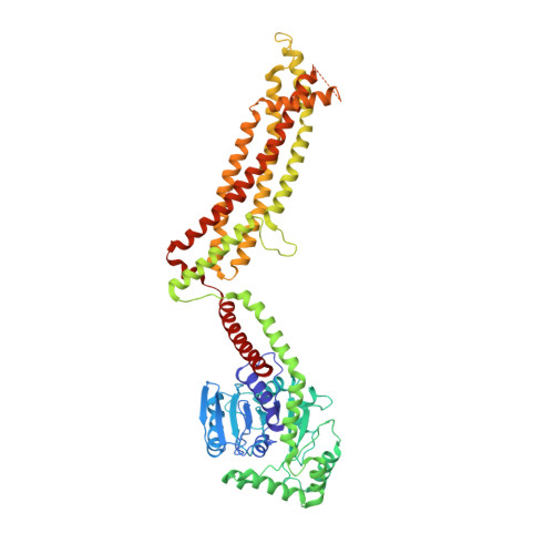
CryoEM structure of MxB reveals a novel oligomerization interface critical for HIV restriction.

Alvarez, F.J.D.He, S.Perilla, J.R.Jang, S.Schulten, K.Engelman, A.N.Scheres, S.H.W.Zhang, P.

(2017) Sci Adv 3: e1701264-e1701264

  • DOI: https://doi.org/10.1126/sciadv.1701264
  • Primary Citation Related Structures: 
    5UOT

  • PubMed Abstract: 

    Human dynamin-like, interferon-induced myxovirus resistance 2 (Mx2 or MxB) is a potent HIV-1 inhibitor. Antiviral activity requires both the amino-terminal region of MxB and protein oligomerization, each of which has eluded structural determination due to difficulties in protein preparation. We report that maltose binding protein-fused, full-length wild-type MxB purifies as oligomers and further self-assembles into helical arrays in physiological salt. Guanosine triphosphate (GTP), but not guanosine diphosphate, binding results in array disassembly, whereas subsequent GTP hydrolysis allows its reformation. Using cryo-electron microscopy (cryoEM), we determined the MxB assembly structure at 4.6 Å resolution, representing the first near-atomic resolution structure in the mammalian dynamin superfamily. The structure revealed previously described and novel MxB assembly interfaces. Mutational analyses demonstrated a critical role for one of the novel interfaces in HIV-1 restriction.


  • Organizational Affiliation
    • Department of Structural Biology, University of Pittsburgh School of Medicine, Pittsburgh, PA 15260, USA.

Macromolecule Content 

  • Total Structure Weight: 4,400.74 kDa 
  • Atom Count: 298,034 
  • Modeled Residue Count: 36,952 
  • Deposited Residue Count: 38,378 
  • Unique protein chains: 1

Macromolecules

Find similar proteins by:|  3D Structure
Entity ID: 1
MoleculeChains  Sequence LengthOrganismDetailsImage
Interferon-induced GTP-binding protein Mx2619Homo sapiensMutation(s): 0 
Gene Names: MX2
UniProt & NIH Common Fund Data Resources
Find proteins for P20592 (Homo sapiens)
Explore P20592 
Go to UniProtKB:  P20592
PHAROS:  P20592
GTEx:  ENSG00000183486 
Entity Groups
Sequence Clusters30% Identity50% Identity70% Identity90% Identity95% Identity100% Identity
UniProt GroupP20592
Sequence Annotations
Expand
Reference Sequence

Experimental Data & Validation

Experimental Data

  • Method: ELECTRON MICROSCOPY
  • Resolution: 4.60 Å
  • Aggregation State: HELICAL ARRAY 
  • Reconstruction Method: HELICAL 
EM Software:
TaskSoftware PackageVersion
MODEL REFINEMENTMDFF3.0
RECONSTRUCTIONRELION2.0 beta

Structure Validation

View Full Validation Report


Entry History 

& Funding Information

Deposition Data


Funding OrganizationLocationGrant Number
National Institutes of HealthUnited StatesGM082251, GM085043, GM104601, GM067887, AI039394
Medical Research Council (United Kingdom)United KingdomMC_UP_A025_1013
National Science FoundationUnited StatesOCI 07-25070, ACI-1238993

Revision History  (Full details and data files)

  • Version 1.0: 2018-02-21
    Type: Initial release
  • Version 1.1: 2018-10-24
    Changes: Data collection, Database references, Other
  • Version 1.2: 2019-01-23
    Changes: Data collection, Refinement description
  • Version 1.3: 2024-05-15
    Changes: Data collection, Database references, Refinement description