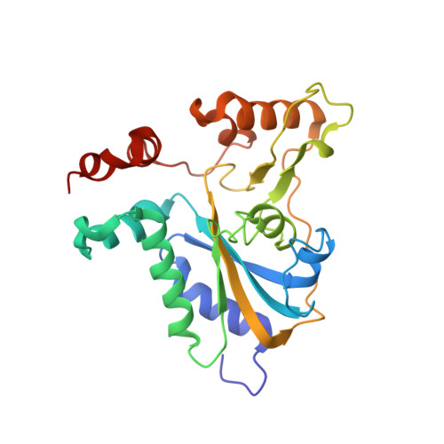
5UOS

Crystal Structure of CblC (MMACHC) (1-238), a human B12 processing enzyme, complexed with an Antivitamin B12


Experimental Data Snapshot

  • Method: X-RAY DIFFRACTION
  • Resolution: 2.51 Å
  • R-Value Free: 0.214 
  • R-Value Work: 0.176 
  • R-Value Observed: 0.178 

Starting Model: experimental
View more details

wwPDB Validation   3D Report Full Report


Ligand Structure Quality Assessment 


This is version 1.4 of the entry. See complete history


Literature

Antivitamin B12 Inhibition of the Human B12 -Processing Enzyme CblC: Crystal Structure of an Inactive Ternary Complex with Glutathione as the Cosubstrate.

Ruetz, M.Shanmuganathan, A.Gherasim, C.Karasik, A.Salchner, R.Kieninger, C.Wurst, K.Banerjee, R.Koutmos, M.Krautler, B.

(2017) Angew Chem Int Ed Engl 56: 7387-7392

  • DOI: https://doi.org/10.1002/anie.201701583
  • Primary Citation of Related Structures:  
    5UOS

  • PubMed Abstract: 

    B 12 antivitamins are important and robust tools for investigating the biological roles of vitamin B 12 . Here, the potential antivitamin B 12 2,4-difluorophenylethynylcobalamin (F2PhEtyCbl) was prepared, and its 3D structure was studied in solution and in the crystal. Chemically inert F2PhEtyCbl resisted thermolysis of its Co-C bond at 100 °C, was stable in bright daylight, and also remained intact upon prolonged storage in aqueous solution at room temperature. It binds to the human B 12 -processing enzyme CblC with high affinity (K D =130 nm) in the presence of the cosubstrate glutathione (GSH). F2PhEtyCbl withstood tailoring by CblC, and it also stabilized the ternary complex with GSH. The crystal structure of this inactivated assembly provides first insight into the binding interactions between an antivitamin B 12 and CblC, as well as into the organization of GSH and a base-off cobalamin in the active site of this enzyme.


  • Organizational Affiliation

    Institute of Organic Chemistry and Center for Molecular Biosciences, University of Innsbruck, 6020, Innsbruck, Austria.


Macromolecules
Find similar proteins by:  (by identity cutoff)  |  3D Structure
Entity ID: 1
MoleculeChains Sequence LengthOrganismDetailsImage
Methylmalonic aciduria and homocystinuria type C protein241Homo sapiensMutation(s): 0 
Gene Names: MMACHC
EC: 1.16.1 (PDB Primary Data), 2.5.1.151 (UniProt), 1.16.1.6 (UniProt)
UniProt & NIH Common Fund Data Resources
Find proteins for Q9Y4U1 (Homo sapiens)
Explore Q9Y4U1 
Go to UniProtKB:  Q9Y4U1
PHAROS:  Q9Y4U1
GTEx:  ENSG00000132763 
Entity Groups  
Sequence Clusters30% Identity50% Identity70% Identity90% Identity95% Identity100% Identity
UniProt GroupQ9Y4U1
Sequence Annotations
Expand
  • Reference Sequence
Small Molecules
Ligands 6 Unique
IDChains Name / Formula / InChI Key2D Diagram3D Interactions
B12
Query on B12

Download Ideal Coordinates CCD File 
B [auth A]COBALAMIN
C62 H89 Co N13 O14 P
LKVIQTCSMMVGFU-DWSMJLPVSA-N
GSH
Query on GSH

Download Ideal Coordinates CCD File 
D [auth A]GLUTATHIONE
C10 H17 N3 O6 S
RWSXRVCMGQZWBV-WDSKDSINSA-N
171
Query on 171

Download Ideal Coordinates CCD File 
E [auth A]2-PHENYLAMINO-ETHANESULFONIC ACID
C8 H11 N O3 S
IAVHKMVGTPXJIC-UHFFFAOYSA-N
8FS
Query on 8FS

Download Ideal Coordinates CCD File 
C [auth A]1-ethynyl-2,4-difluorobenzene
C8 H4 F2
HRUJQXRGWQWYDH-UHFFFAOYSA-N
SO4
Query on SO4

Download Ideal Coordinates CCD File 
F [auth A],
G [auth A]
SULFATE ION
O4 S
QAOWNCQODCNURD-UHFFFAOYSA-L
NA
Query on NA

Download Ideal Coordinates CCD File 
H [auth A]SODIUM ION
Na
FKNQFGJONOIPTF-UHFFFAOYSA-N
Experimental Data & Validation

Experimental Data

  • Method: X-RAY DIFFRACTION
  • Resolution: 2.51 Å
  • R-Value Free: 0.214 
  • R-Value Work: 0.176 
  • R-Value Observed: 0.178 
  • Space Group: P 61 2 2
Unit Cell:
Length ( Å )Angle ( ˚ )
a = 114.432α = 90
b = 114.432β = 90
c = 150.631γ = 120
Software Package:
Software NamePurpose
REFMACrefinement
XDSdata reduction
Aimlessdata scaling
PHASERphasing

Structure Validation

View Full Validation Report


Ligand Structure Quality Assessment 


Entry History & Funding Information

Deposition Data


Funding OrganizationLocationGrant Number
American Heart AssociationUnited States13SDG14560056

Revision History  (Full details and data files)

  • Version 1.0: 2017-06-07
    Type: Initial release
  • Version 1.1: 2017-06-21
    Changes: Database references
  • Version 1.2: 2017-09-27
    Changes: Author supporting evidence
  • Version 1.3: 2017-11-01
    Changes: Author supporting evidence
  • Version 1.4: 2023-10-04
    Changes: Data collection, Database references, Refinement description