5UN8 | pdb_00005un8

Crystal Structure of human O-GlcNAcase in complex with glycopeptide p53


Experimental Data Snapshot

  • Method: X-RAY DIFFRACTION
  • Resolution: 2.13 Å
  • R-Value Free: 
    0.229 (Depositor), 0.230 (DCC) 
  • R-Value Work: 
    0.184 (Depositor), 0.185 (DCC) 
  • R-Value Observed: 
    0.187 (Depositor) 

Starting Model: experimental
View more details

wwPDB Validation 3D Report Full Report

Validation slider image for 5UN8

Ligand Structure Quality Assessment 


This is version 1.5 of the entry. See complete history

Literature

Structures of human O-GlcNAcase and its complexes reveal a new substrate recognition mode.

Li, B.Li, H.Lu, L.Jiang, J.

(2017) Nat Struct Mol Biol 24: 362-369

  • DOI: https://doi.org/10.1038/nsmb.3390
  • Primary Citation Related Structures: 
    5TKE, 5UN8, 5UN9

  • PubMed Abstract: 

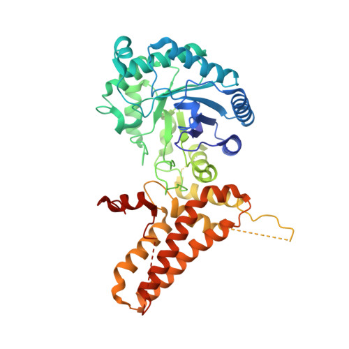
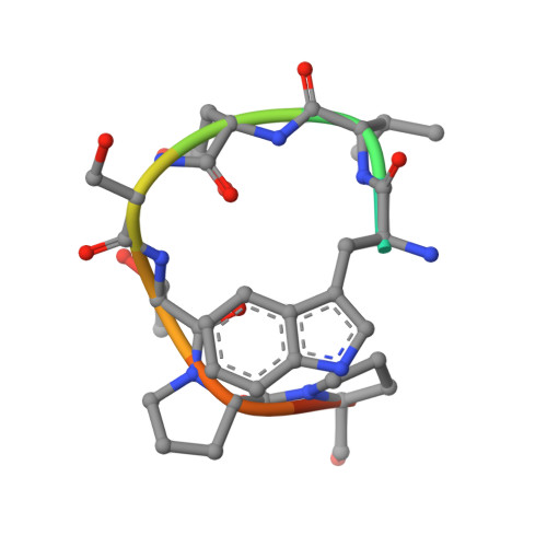
    Human O-GlcNAcase (hOGA) is the unique enzyme responsible for the hydrolysis of the O-linked β-N-acetyl glucosamine (O-GlcNAc) modification, an essential protein glycosylation event that modulates the function of numerous cellular proteins in response to nutrients and stress. Here we report crystal structures of a truncated hOGA, which comprises the catalytic and stalk domains, in apo form, in complex with an inhibitor, and in complex with a glycopeptide substrate. We found that hOGA forms an unusual arm-in-arm homodimer in which the catalytic domain of one monomer is covered by the stalk domain of the sister monomer to create a substrate-binding cleft. Notably, the residues on the cleft surface afford extensive interactions with the peptide substrate in a recognition mode that is distinct from that of its bacterial homologs. These structures represent the first model of eukaryotic enzymes in the glycoside hydrolase 84 (GH84) family and provide a crucial starting point for understanding the substrate specificity of hOGA, which regulates a broad range of biological and pathological processes.


  • Organizational Affiliation
    • Pharmaceutical Sciences Division, School of Pharmacy, University of Wisconsin-Madison, Madison, Wisconsin, USA.

Macromolecule Content 

  • Total Structure Weight: 236.96 kDa 
  • Atom Count: 15,552 
  • Modeled Residue Count: 1,740 
  • Deposited Residue Count: 2,060 
  • Unique protein chains: 2

Macromolecules

Find similar proteins by:|  3D Structure
Entity ID: 1
MoleculeChains  Sequence LengthOrganismDetailsImage
Protein O-GlcNAcase
A, B, C, D
504Homo sapiensMutation(s): 0 
Gene Names: MGEA5HEXCKIAA0679MEA5
EC: 3.2.1.169 (PDB Primary Data), 3.2.1 (PDB Primary Data)
UniProt & NIH Common Fund Data Resources
Find proteins for O60502 (Homo sapiens)
Explore O60502 
Go to UniProtKB:  O60502
PHAROS:  O60502
GTEx:  ENSG00000198408 
Entity Groups
Sequence Clusters30% Identity50% Identity70% Identity90% Identity95% Identity100% Identity
UniProt GroupO60502
Sequence Annotations
Expand
Reference Sequence
Find similar proteins by:  Sequence   |   3D Structure  
Entity ID: 2
MoleculeChains  Sequence LengthOrganismDetailsImage
P53 peptide
E, F, G, H
11Homo sapiensMutation(s): 0 
UniProt & NIH Common Fund Data Resources
Find proteins for P04637 (Homo sapiens)
Explore P04637 
Go to UniProtKB:  P04637
PHAROS:  P04637
GTEx:  ENSG00000141510 
Entity Groups
Sequence Clusters30% Identity50% Identity70% Identity90% Identity95% Identity100% Identity
UniProt GroupP04637
Glycosylation
Glycosylation Sites: 1Go to GlyGen: P04637-1
Sequence Annotations
Expand
Reference Sequence

Experimental Data & Validation

Experimental Data

  • Method: X-RAY DIFFRACTION
  • Resolution: 2.13 Å
  • R-Value Free:  0.229 (Depositor), 0.230 (DCC) 
  • R-Value Work:  0.184 (Depositor), 0.185 (DCC) 
  • R-Value Observed: 0.187 (Depositor) 
Space Group: P 1 21 1
Unit Cell:
Length ( Å )Angle ( ˚ )
a = 89.909α = 90
b = 95.393β = 96.91
c = 149.32γ = 90
Software Package:
Software NamePurpose
REFMACrefinement
HKL-2000data reduction
HKL-2000data scaling
PHASERphasing

Structure Validation

View Full Validation Report


Ligand Structure Quality Assessment 


Entry History 

& Funding Information

Deposition Data


Funding OrganizationLocationGrant Number
University of Wisconsin-MadisonUnited States--

Revision History  (Full details and data files)

  • Version 1.0: 2017-03-15
    Type: Initial release
  • Version 1.1: 2017-04-05
    Changes: Database references
  • Version 1.2: 2017-04-19
    Changes: Database references
  • Version 1.3: 2020-07-29
    Type: Remediation
    Reason: Carbohydrate remediation
    Changes: Data collection, Derived calculations, Structure summary
  • Version 1.4: 2023-10-04
    Changes: Data collection, Database references, Refinement description, Structure summary
  • Version 1.5: 2024-11-06
    Changes: Structure summary