5ULM | pdb_00005ulm

Structure of the ASK1 central regulatory region


Experimental Data Snapshot

  • Method: X-RAY DIFFRACTION
  • Resolution: 2.10 Å
  • R-Value Free: 
    0.267 (Depositor), 0.279 (DCC) 
  • R-Value Work: 
    0.233 (Depositor), 0.242 (DCC) 
  • R-Value Observed: 
    0.235 (Depositor) 

wwPDB Validation 3D Report Full Report

Validation slider image for 5ULM

This is version 1.5 of the entry. See complete history

Literature

Structural basis of autoregulatory scaffolding by apoptosis signal-regulating kinase 1.

Weijman, J.F.Kumar, A.Jamieson, S.A.King, C.M.Caradoc-Davies, T.T.Ledgerwood, E.C.Murphy, J.M.Mace, P.D.

(2017) Proc Natl Acad Sci U S A 114: E2096-E2105

  • DOI: https://doi.org/10.1073/pnas.1620813114
  • Primary Citation Related Structures: 
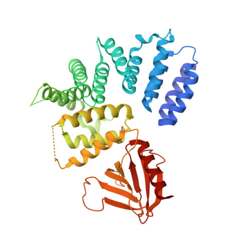
    5ULM

  • PubMed Abstract: 

    Apoptosis signal-regulating kinases (ASK1-3) are apical kinases of the p38 and JNK MAP kinase pathways. They are activated by diverse stress stimuli, including reactive oxygen species, cytokines, and osmotic stress; however, a molecular understanding of how ASK proteins are controlled remains obscure. Here, we report a biochemical analysis of the ASK1 kinase domain in conjunction with its N-terminal thioredoxin-binding domain, along with a central regulatory region that links the two. We show that in solution the central regulatory region mediates a compact arrangement of the kinase and thioredoxin-binding domains and the central regulatory region actively primes MKK6, a key ASK1 substrate, for phosphorylation. The crystal structure of the central regulatory region reveals an unusually compact tetratricopeptide repeat (TPR) region capped by a cryptic pleckstrin homology domain. Biochemical assays show that both a conserved surface on the pleckstrin homology domain and an intact TPR region are required for ASK1 activity. We propose a model in which the central regulatory region promotes ASK1 activity via its pleckstrin homology domain but also facilitates ASK1 autoinhibition by bringing the thioredoxin-binding and kinase domains into close proximity. Such an architecture provides a mechanism for control of ASK-type kinases by diverse activators and inhibitors and demonstrates an unexpected level of autoregulatory scaffolding in mammalian stress-activated MAP kinase signaling.


  • Organizational Affiliation
    • Biochemistry Department, School of Biomedical Sciences, University of Otago, Dunedin 9054, New Zealand.

Macromolecule Content 

  • Total Structure Weight: 90.74 kDa 
  • Atom Count: 6,450 
  • Modeled Residue Count: 749 
  • Deposited Residue Count: 786 
  • Unique protein chains: 1

Macromolecules

Find similar proteins by:|  3D Structure
Entity ID: 1
MoleculeChains  Sequence LengthOrganismDetailsImage
Mitogen-activated protein kinase kinase kinase 5
A, B
393Homo sapiensMutation(s): 0 
Gene Names: MAP3K5ASK1MAPKKK5MEKK5
EC: 2.7.11.25
UniProt & NIH Common Fund Data Resources
Find proteins for Q99683 (Homo sapiens)
Explore Q99683 
Go to UniProtKB:  Q99683
PHAROS:  Q99683
GTEx:  ENSG00000197442 
Entity Groups
Sequence Clusters30% Identity50% Identity70% Identity90% Identity95% Identity100% Identity
UniProt GroupQ99683
Sequence Annotations
Expand
Reference Sequence

Small Molecules

Ligands 1 Unique
IDChains Name / Formula / InChI Key2D Diagram3D Interactions
GOL

Query on GOL


Download:Ideal Coordinates CCD File
C [auth B]GLYCEROL
C3 H8 O3
PEDCQBHIVMGVHV-UHFFFAOYSA-N

Experimental Data & Validation

Experimental Data

  • Method: X-RAY DIFFRACTION
  • Resolution: 2.10 Å
  • R-Value Free:  0.267 (Depositor), 0.279 (DCC) 
  • R-Value Work:  0.233 (Depositor), 0.242 (DCC) 
  • R-Value Observed: 0.235 (Depositor) 
Space Group: P 1 21 1
Unit Cell:
Length ( Å )Angle ( ˚ )
a = 74.234α = 90
b = 57.117β = 104.87
c = 103.575γ = 90
Software Package:
Software NamePurpose
REFMACrefinement
XDSdata reduction
SCALAdata scaling
PHENIXphasing

Structure Validation

View Full Validation Report


Entry History 

Deposition Data

Revision History  (Full details and data files)

  • Version 1.0: 2017-03-01
    Type: Initial release
  • Version 1.1: 2017-03-15
    Changes: Database references
  • Version 1.2: 2017-03-29
    Changes: Database references
  • Version 1.3: 2017-09-27
    Changes: Author supporting evidence, Data collection
  • Version 1.4: 2017-11-01
    Changes: Author supporting evidence
  • Version 1.5: 2024-03-06
    Changes: Data collection, Database references, Refinement description