5UK6 | pdb_00005uk6

Structure of Anabaena Sensory Rhodopsin Determined by Solid State NMR Spectroscopy and DEER


Experimental Data Snapshot

  • Method: SOLID-STATE NMR
  • Conformers Submitted: 10 

wwPDB Validation 3D Report Full Report

Validation slider image for 5UK6

This is version 1.5 of the entry. See complete history

Literature

Oligomeric Structure of Anabaena Sensory Rhodopsin in a Lipid Bilayer Environment by Combining Solid-State NMR and Long-range DEER Constraints.

Milikisiyants, S.Wang, S.Munro, R.A.Donohue, M.Ward, M.E.Bolton, D.Brown, L.S.Smirnova, T.I.Ladizhansky, V.Smirnov, A.I.

(2017) J Mol Biology 429: 1903-1920

  • DOI: https://doi.org/10.1016/j.jmb.2017.05.005
  • Primary Citation Related Structures: 
    5UK6

  • PubMed Abstract: 

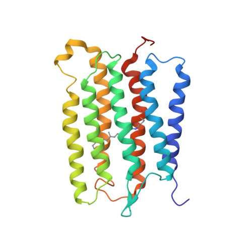
    Oligomerization of membrane proteins is common in nature. Here, we combine spin-labeling double electron-electron resonance (DEER) and solid-state NMR (ssNMR) spectroscopy to refine the structure of an oligomeric integral membrane protein, Anabaena sensory rhodopsin (ASR), reconstituted in a lipid environment. An essential feature of such a combined approach is that it provides structural distance restraints spanning a range of ca 3-60Å while using the same sample preparation (i.e., mutations, paramagnetic labeling, and reconstitution in lipid bilayers) for both ssNMR and DEER. Direct modeling of the multispin effects on DEER signal allowed for the determination of the oligomeric order and for obtaining long-range DEER distance restraints between the ASR trimer subunits that were used to refine the ssNMR structure of ASR. The improved structure of the ASR trimer revealed a more compact packing of helices and side chains at the intermonomer interface, compared to the structure determined using the ssNMR data alone. The extent of the refinement is significant when compared with typical helix movements observed for the active states of homologous proteins. Our combined approach of using complementary DEER and NMR measurements for the determination of oligomeric structures would be widely applicable to membrane proteins where paramagnetic tags can be introduced. Such a method could be used to study the effects of the lipid membrane composition on protein oligomerization and to observe structural changes in protein oligomers upon drug, substrate, and co-factor binding.


  • Organizational Affiliation
    • Department of Chemistry, College of Sciences, North Carolina State University, 2620 Yarbrough Dive, Raleigh, NC 27695-8204, USA.

Macromolecule Content 

  • Total Structure Weight: 82.54 kDa 
  • Atom Count: 5,664 
  • Modeled Residue Count: 687 
  • Deposited Residue Count: 705 
  • Unique protein chains: 1

Macromolecules

Find similar proteins by:|  3D Structure
Entity ID: 1
MoleculeChains  Sequence LengthOrganismDetailsImage
Bacteriorhodopsin
A, B, C
235Nostoc sp. PCC 7120 = FACHB-418Mutation(s): 0 
Gene Names: alr3165
UniProt
Find proteins for A0ACD6B968 (Nostoc sp. (strain PCC 7120 / SAG 25.82 / UTEX 2576))
Explore A0ACD6B968 
Go to UniProtKB:  A0ACD6B968
Entity Groups
Sequence Clusters30% Identity50% Identity70% Identity90% Identity95% Identity100% Identity
UniProt GroupA0ACD6B968
Sequence Annotations
Expand
Reference Sequence

Small Molecules

Modified Residues  1 Unique
IDChains TypeFormula2D DiagramParent
LYR
Query on LYR
A, B, C
L-PEPTIDE LINKINGC26 H42 N2 O2LYS

Experimental Data & Validation

Experimental Data

  • Method: SOLID-STATE NMR
  • Conformers Submitted: 10 

Structure Validation

View Full Validation Report


Entry History 

& Funding Information

Deposition Data


Funding OrganizationLocationGrant Number
Department of Energy (DOE, United States)United StatesDE-FG02-02ER15354
Natural Sciences and Engineering Research Council (NSERC, Canada)CanadaRGPIN-2014-04547
National Natural Science Foundation of China (NSFC)China31470727
National Science Foundation (NSF, United States)United StatesDBI-1229547
Ministry of Science and Technology (MoST, China)China2016YFA0501203

Revision History  (Full details and data files)

  • Version 1.0: 2017-05-31
    Type: Initial release
  • Version 1.1: 2017-06-21
    Changes: Database references
  • Version 1.2: 2017-09-27
    Changes: Author supporting evidence
  • Version 1.3: 2019-11-27
    Changes: Author supporting evidence, Data collection
  • Version 1.4: 2020-01-08
    Changes: Author supporting evidence
  • Version 1.5: 2025-04-02
    Changes: Data collection, Database references, Structure summary