5UFP | pdb_00005ufp

Crystal structure of PT2399 bound to HIF2a-B*:ARNT-B* complex


Experimental Data Snapshot

  • Method: X-RAY DIFFRACTION
  • Resolution: 1.90 Å
  • R-Value Free: 
    0.271 (Depositor), 0.272 (DCC) 
  • R-Value Work: 
    0.218 (Depositor), 0.219 (DCC) 
  • R-Value Observed: 
    0.221 (Depositor) 

wwPDB Validation 3D Report Full Report

Validation slider image for 5UFP

Ligand Structure Quality Assessment 


This is version 1.2 of the entry. See complete history

Literature

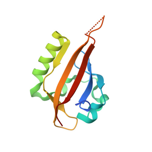
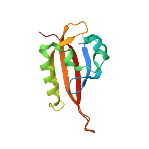
On-target efficacy of a HIF-2 alpha antagonist in preclinical kidney cancer models.

Cho, H.Du, X.Rizzi, J.P.Liberzon, E.Chakraborty, A.A.Gao, W.Carvo, I.Signoretti, S.Bruick, R.K.Josey, J.A.Wallace, E.M.Kaelin, W.G.

(2016) Nature 539: 107-111

  • DOI: https://doi.org/10.1038/nature19795
  • Primary Citation Related Structures: 
    5UFP

  • PubMed Abstract: 

    Clear cell renal cell carcinoma, the most common form of kidney cancer, is usually linked to inactivation of the pVHL tumour suppressor protein and consequent accumulation of the HIF-2α transcription factor (also known as EPAS1). Here we show that a small molecule (PT2399) that directly inhibits HIF-2α causes tumour regression in preclinical mouse models of primary and metastatic pVHL-defective clear cell renal cell carcinoma in an on-target fashion. pVHL-defective clear cell renal cell carcinoma cell lines display unexpectedly variable sensitivity to PT2399, however, suggesting the need for predictive biomarkers to be developed to use this approach optimally in the clinic.


  • Organizational Affiliation
    • Department of Medical Oncology, Dana-Farber Cancer Institute and Brigham and Women's Hospital, Harvard Medical School, Boston, Massachusetts 02215, USA.

Macromolecule Content 

  • Total Structure Weight: 27.47 kDa 
  • Atom Count: 1,853 
  • Modeled Residue Count: 214 
  • Deposited Residue Count: 232 
  • Unique protein chains: 2

Macromolecules

Find similar proteins by:|  3D Structure
Entity ID: 1
MoleculeChains  Sequence LengthOrganismDetailsImage
Endothelial PAS domain-containing protein 1115Homo sapiensMutation(s): 1 
Gene Names: EPAS1BHLHE73HIF2AMOP2PASD2
UniProt & NIH Common Fund Data Resources
Find proteins for Q99814 (Homo sapiens)
Explore Q99814 
Go to UniProtKB:  Q99814
PHAROS:  Q99814
GTEx:  ENSG00000116016 
Entity Groups
Sequence Clusters30% Identity50% Identity70% Identity90% Identity95% Identity100% Identity
UniProt GroupQ99814
Sequence Annotations
Expand
Reference Sequence
Find similar proteins by:|  3D Structure
Entity ID: 2
MoleculeChains  Sequence LengthOrganismDetailsImage
Aryl hydrocarbon receptor nuclear translocator117Homo sapiensMutation(s): 1 
Gene Names: ARNTBHLHE2
UniProt & NIH Common Fund Data Resources
Find proteins for P27540 (Homo sapiens)
Explore P27540 
Go to UniProtKB:  P27540
PHAROS:  P27540
GTEx:  ENSG00000143437 
Entity Groups
Sequence Clusters30% Identity50% Identity70% Identity90% Identity95% Identity100% Identity
UniProt GroupP27540
Sequence Annotations
Expand
Reference Sequence

Small Molecules

Ligands 1 Unique
IDChains Name / Formula / InChI Key2D Diagram3D Interactions
86D

Query on 86D


Download:Ideal Coordinates CCD File
C [auth A]3-({(1S)-7-[(difluoromethyl)sulfonyl]-2,2-difluoro-1-hydroxy-2,3-dihydro-1H-inden-4-yl}oxy)-5-fluorobenzonitrile
C17 H10 F5 N O4 S
MXUSGDMIHGLCNC-HNNXBMFYSA-N
Binding Affinity Annotations 
IDSourceBinding Affinity
86D BindingDB:  5UFP IC50: min: 5, max: 8 (nM) from 2 assay(s)
EC50: min: 6, max: 233 (nM) from 3 assay(s)

Experimental Data & Validation

Experimental Data

  • Method: X-RAY DIFFRACTION
  • Resolution: 1.90 Å
  • R-Value Free:  0.271 (Depositor), 0.272 (DCC) 
  • R-Value Work:  0.218 (Depositor), 0.219 (DCC) 
  • R-Value Observed: 0.221 (Depositor) 
Space Group: C 1 2 1
Unit Cell:
Length ( Å )Angle ( ˚ )
a = 73.379α = 90
b = 84.057β = 106.48
c = 41.472γ = 90
Software Package:
Software NamePurpose
REFMACrefinement
HKL-2000data collection
SCALEPACKdata scaling
PHASERphasing
PDB_EXTRACTdata extraction
HKL-2000data reduction
DENZOdata reduction

Structure Validation

View Full Validation Report


Ligand Structure Quality Assessment 


Entry History 

Deposition Data

  • Released Date: 2017-01-25 
  • Deposition Author(s): Du, X.
  • This entry supersedes: 5T0T

Revision History  (Full details and data files)

  • Version 1.0: 2017-01-25
    Type: Initial release
  • Version 1.1: 2017-11-22
    Changes: Refinement description
  • Version 1.2: 2024-03-06
    Changes: Data collection, Database references