5U74

Structure of human Niemann-Pick C1 protein


Experimental Data Snapshot

  • Method: X-RAY DIFFRACTION
  • Resolution: 3.33 Å
  • R-Value Free: 0.299 
  • R-Value Work: 0.246 
  • R-Value Observed: 0.249 

wwPDB Validation   3D Report Full Report


Ligand Structure Quality Assessment 


This is version 2.1 of the entry. See complete history


Literature

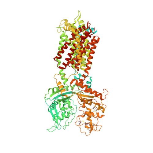
3.3 angstrom structure of Niemann-Pick C1 protein reveals insights into the function of the C-terminal luminal domain in cholesterol transport.

Li, X.Lu, F.Trinh, M.N.Schmiege, P.Seemann, J.Wang, J.Blobel, G.

(2017) Proc Natl Acad Sci U S A 114: 9116-9121

  • DOI: https://doi.org/10.1073/pnas.1711716114
  • Primary Citation of Related Structures:  
    5U73, 5U74

  • PubMed Abstract: 

    Niemann-Pick C1 (NPC1) and NPC2 proteins are indispensable for the export of LDL-derived cholesterol from late endosomes. Mutations in these proteins result in Niemann-Pick type C disease, a lysosomal storage disease. Despite recent reports of the NPC1 structure depicting its overall architecture, the function of its C-terminal luminal domain (CTD) remains poorly understood even though 45% of NPC disease-causing mutations are in this domain. Here, we report a crystal structure at 3.3 Å resolution of NPC1* (residues 314-1,278), which-in contrast to previous lower resolution structures-features the entire CTD well resolved. Notably, all eight cysteines of the CTD form four disulfide bonds, one of which (C909-C914) enforces a specific loop that in turn mediates an interaction with a loop of the N-terminal domain (NTD). Importantly, this loop and its interaction with the NTD were not observed in any previous structures due to the lower resolution. Our mutagenesis experiments highlight the physiological relevance of the CTD-NTD interaction, which might function to keep the NTD in the proper orientation for receiving cholesterol from NPC2. Additionally, this structure allows us to more precisely map all of the disease-causing mutations, allowing future molecular insights into the pathogenesis of NPC disease.


  • Organizational Affiliation

    Laboratory of Cell Biology, The Rockefeller University, New York, NY 10065; xli05@rockefeller.edu blobel@rockefeller.edu.


Macromolecules
Find similar proteins by:  (by identity cutoff)  |  3D Structure
Entity ID: 1
MoleculeChains Sequence LengthOrganismDetailsImage
Niemann-Pick C1 protein1,278Homo sapiensMutation(s): 0 
Gene Names: NPC1
Membrane Entity: Yes 
UniProt & NIH Common Fund Data Resources
Find proteins for O15118 (Homo sapiens)
Explore O15118 
Go to UniProtKB:  O15118
PHAROS:  O15118
GTEx:  ENSG00000141458 
Entity Groups  
Sequence Clusters30% Identity50% Identity70% Identity90% Identity95% Identity100% Identity
UniProt GroupO15118
Sequence Annotations
Expand
  • Reference Sequence
Oligosaccharides

Help

Entity ID: 2
MoleculeChains Length2D Diagram Glycosylation3D Interactions
2-acetamido-2-deoxy-beta-D-glucopyranose-(1-4)-2-acetamido-2-deoxy-beta-D-glucopyranose
B
2N-Glycosylation
Glycosylation Resources
GlyTouCan:  G42666HT
GlyCosmos:  G42666HT
GlyGen:  G42666HT
Entity ID: 3
MoleculeChains Length2D Diagram Glycosylation3D Interactions
alpha-D-mannopyranose-(1-3)-2-acetamido-2-deoxy-beta-D-glucopyranose-(1-4)-2-acetamido-2-deoxy-beta-D-glucopyranose
C
3N-Glycosylation
Glycosylation Resources
GlyTouCan:  G66019PC
GlyCosmos:  G66019PC
GlyGen:  G66019PC
Entity ID: 4
MoleculeChains Length2D Diagram Glycosylation3D Interactions
alpha-D-mannopyranose-(1-4)-2-acetamido-2-deoxy-beta-D-glucopyranose-(1-4)-2-acetamido-2-deoxy-beta-D-glucopyranose
D
3N-Glycosylation
Glycosylation Resources
GlyTouCan:  G62182OO
GlyCosmos:  G62182OO
GlyGen:  G62182OO
Small Molecules
Ligands 2 Unique
IDChains Name / Formula / InChI Key2D Diagram3D Interactions
NAG
Query on NAG

Download Ideal Coordinates CCD File 
E [auth A]
F [auth A]
G [auth A]
H [auth A]
I [auth A]
E [auth A],
F [auth A],
G [auth A],
H [auth A],
I [auth A],
J [auth A],
K [auth A],
L [auth A]
2-acetamido-2-deoxy-beta-D-glucopyranose
C8 H15 N O6
OVRNDRQMDRJTHS-FMDGEEDCSA-N
MES
Query on MES

Download Ideal Coordinates CCD File 
M [auth A]2-(N-MORPHOLINO)-ETHANESULFONIC ACID
C6 H13 N O4 S
SXGZJKUKBWWHRA-UHFFFAOYSA-N
Experimental Data & Validation

Experimental Data

  • Method: X-RAY DIFFRACTION
  • Resolution: 3.33 Å
  • R-Value Free: 0.299 
  • R-Value Work: 0.246 
  • R-Value Observed: 0.249 
  • Space Group: C 2 2 21
Unit Cell:
Length ( Å )Angle ( ˚ )
a = 174.87α = 90
b = 222.115β = 90
c = 107.851γ = 90
Software Package:
Software NamePurpose
PHENIXrefinement
HKL-2000data reduction
HKL-2000data scaling
PHASERphasing

Structure Validation

View Full Validation Report


Ligand Structure Quality Assessment 


Entry History 

Deposition Data

  • Released Date: 2017-10-11 
  • Deposition Author(s): Li, X.

Revision History  (Full details and data files)

  • Version 1.0: 2017-10-11
    Type: Initial release
  • Version 2.0: 2020-07-29
    Type: Remediation
    Reason: Carbohydrate remediation
    Changes: Atomic model, Data collection, Derived calculations, Structure summary
  • Version 2.1: 2023-10-04
    Changes: Data collection, Database references, Refinement description, Structure summary