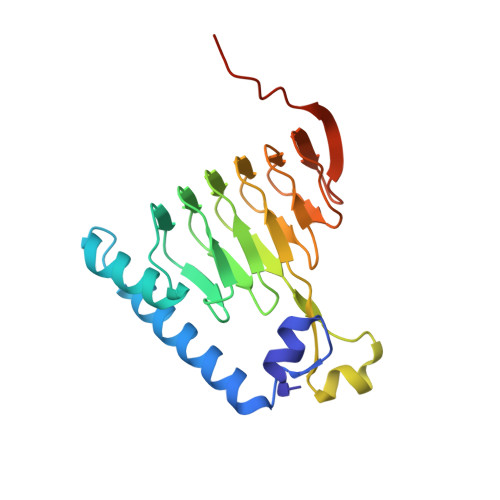
Crystal structure of Galactoside O-acetyltransferase complex with CoA (H3 space group)
Czub, M.P., Porebski, P.J., Knapik, A.A., Niedzialkowska, E., Siuda, M.K., Anderson, W.F., Minor, W., Center for Structural Genomics of Infectious DiseasesTo be published.