5U2J | pdb_00005u2j

MORF double PHD finger (DPF) in complex with histone H3K14bu


Experimental Data Snapshot

  • Method: X-RAY DIFFRACTION
  • Resolution: 1.60 Å
  • R-Value Free: 
    0.238 (Depositor), 0.241 (DCC) 
  • R-Value Work: 
    0.191 (Depositor), 0.193 (DCC) 
  • R-Value Observed: 
    0.193 (Depositor) 

wwPDB Validation 3D Report Full Report

Validation slider image for 5U2J

This is version 1.2 of the entry. See complete history

Literature

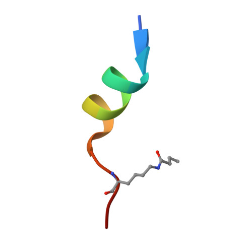
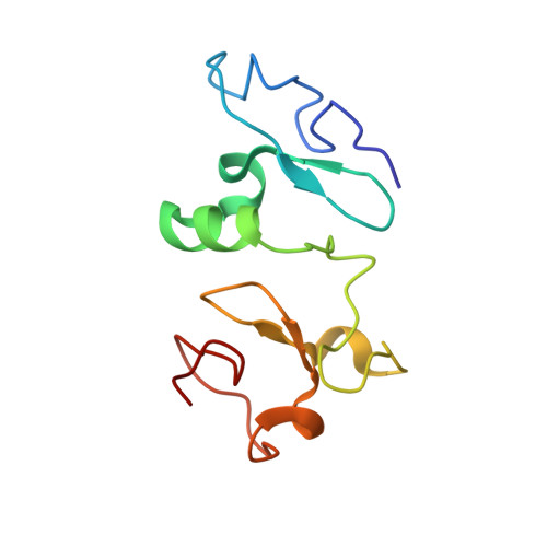
Recognition of Histone H3K14 Acylation by MORF.

Klein, B.J.Simithy, J.Wang, X.Ahn, J.Andrews, F.H.Zhang, Y.Cote, J.Shi, X.Garcia, B.A.Kutateladze, T.G.

(2017) Structure 25: 650-654.e2

  • DOI: https://doi.org/10.1016/j.str.2017.02.003
  • Primary Citation Related Structures: 
    5U2J

  • PubMed Abstract: 

    The monocytic leukemia zinc-finger protein-related factor (MORF) is a transcriptional coactivator and a catalytic subunit of the lysine acetyltransferase complex implicated in cancer and developmental diseases. We have previously shown that the double plant homeodomain finger (DPF) of MORF is capable of binding to acetylated histone H3. Here we demonstrate that the DPF of MORF recognizes many newly identified acylation marks. The mass spectrometry study provides comprehensive analysis of H3K14 acylation states in vitro and in vivo. The crystal structure of the MORF DPF-H3K14butyryl complex offers insight into the selectivity of this reader toward lipophilic acyllysine substrates. Together, our findings support the mechanism by which the acetyltransferase MORF promotes spreading of histone acylation.


  • Organizational Affiliation
    • Department of Pharmacology, University of Colorado School of Medicine, Aurora, CO 80045, USA.

Macromolecule Content 

  • Total Structure Weight: 28.94 kDa 
  • Atom Count: 2,221 
  • Modeled Residue Count: 253 
  • Deposited Residue Count: 254 
  • Unique protein chains: 2

Macromolecules

Find similar proteins by:  Sequence   |   3D Structure  
Entity ID: 1
MoleculeChains  Sequence LengthOrganismDetailsImage
Histone H3K14buA [auth D],
D [auth C]
16Homo sapiensMutation(s): 0 
UniProt & NIH Common Fund Data Resources
Find proteins for P68431 (Homo sapiens)
Explore P68431 
Go to UniProtKB:  P68431
Entity Groups
Sequence Clusters30% Identity50% Identity70% Identity90% Identity95% Identity100% Identity
UniProt GroupP68431
Sequence Annotations
Expand
Reference Sequence
Find similar proteins by:|  3D Structure
Entity ID: 2
MoleculeChains  Sequence LengthOrganismDetailsImage
Histone acetyltransferase KAT6BB [auth A],
C [auth B]
111Homo sapiensMutation(s): 0 
Gene Names: KAT6BKIAA0383MORFMOZ2MYST4
EC: 2.3.1.48
UniProt & NIH Common Fund Data Resources
Find proteins for Q8WYB5 (Homo sapiens)
Explore Q8WYB5 
Go to UniProtKB:  Q8WYB5
PHAROS:  Q8WYB5
GTEx:  ENSG00000156650 
Entity Groups
Sequence Clusters30% Identity50% Identity70% Identity90% Identity95% Identity100% Identity
UniProt GroupQ8WYB5
Sequence Annotations
Expand
Reference Sequence

Small Molecules

Modified Residues  1 Unique
IDChains TypeFormula2D DiagramParent
BTK
Query on BTK
A [auth D],
D [auth C]
L-PEPTIDE LINKINGC10 H20 N2 O3LYS

Experimental Data & Validation

Experimental Data

  • Method: X-RAY DIFFRACTION
  • Resolution: 1.60 Å
  • R-Value Free:  0.238 (Depositor), 0.241 (DCC) 
  • R-Value Work:  0.191 (Depositor), 0.193 (DCC) 
  • R-Value Observed: 0.193 (Depositor) 
Space Group: P 1 21 1
Unit Cell:
Length ( Å )Angle ( ˚ )
a = 32.54α = 90
b = 69.19β = 97.16
c = 69.93γ = 90
Software Package:
Software NamePurpose
PHENIXrefinement
XDSdata reduction
SCALAdata scaling
PHASERphasing

Structure Validation

View Full Validation Report


Entry History 

Deposition Data

Revision History  (Full details and data files)

  • Version 1.0: 2017-04-12
    Type: Initial release
  • Version 1.1: 2017-04-19
    Changes: Database references
  • Version 1.2: 2025-04-02
    Changes: Data collection, Database references, Derived calculations, Structure summary