5TY4 | pdb_00005ty4

MicroED structure of a complex between monomeric TGF-b and its receptor, TbRII, at 2.9 A resolution


Experimental Data Snapshot

  • Method: ELECTRON CRYSTALLOGRAPHY
  • Resolution: 2.90 Å
  • R-Value Free: 
    0.328 (Depositor) 
  • R-Value Work: 
    0.292 (Depositor) 
  • R-Value Observed: 
    0.295 (Depositor) 

Starting Model: experimental
View more details

wwPDB Validation 3D Report Full Report

Validation slider image for 5TY4

This is version 1.5 of the entry. See complete history

Literature

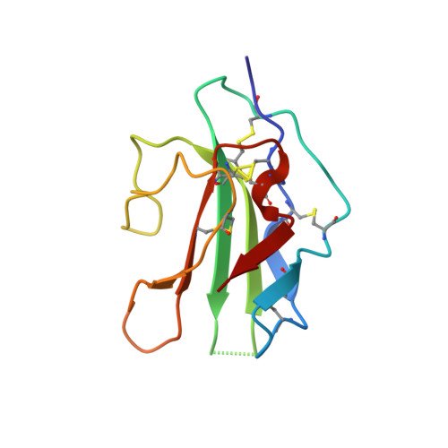
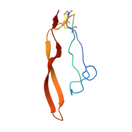
Atomic-resolution structures from fragmented protein crystals with the cryoEM method MicroED.

de la Cruz, M.J.Hattne, J.Shi, D.Seidler, P.Rodriguez, J.Reyes, F.E.Sawaya, M.R.Cascio, D.Weiss, S.C.Kim, S.K.Hinck, C.S.Hinck, A.P.Calero, G.Eisenberg, D.Gonen, T.

(2017) Nat Methods 14: 399-402

  • DOI: https://doi.org/10.1038/nmeth.4178
  • Primary Citation Related Structures: 
    5K7N, 5K7O, 5K7P, 5K7Q, 5K7R, 5K7S, 5K7T, 5TY4

  • PubMed Abstract: 

    Traditionally, crystallographic analysis of macromolecules has depended on large, well-ordered crystals, which often require significant effort to obtain. Even sizable crystals sometimes suffer from pathologies that render them inappropriate for high-resolution structure determination. Here we show that fragmentation of large, imperfect crystals into microcrystals or nanocrystals can provide a simple path for high-resolution structure determination by the cryoEM method MicroED and potentially by serial femtosecond crystallography.


  • Organizational Affiliation
    • Howard Hughes Medical Institute, Janelia Research Campus, Ashburn, Virginia, USA.

Macromolecule Content 

  • Total Structure Weight: 22.87 kDa 
  • Atom Count: 1,327 
  • Modeled Residue Count: 166 
  • Deposited Residue Count: 200 
  • Unique protein chains: 2

Macromolecules

Find similar proteins by:|  3D Structure
Entity ID: 1
MoleculeChains  Sequence LengthOrganismDetailsImage
TGF-beta receptor type-2103Homo sapiensMutation(s): 0 
Gene Names: TGFBR2
EC: 2.7.11.30
UniProt & NIH Common Fund Data Resources
Find proteins for P37173 (Homo sapiens)
Explore P37173 
Go to UniProtKB:  P37173
PHAROS:  P37173
GTEx:  ENSG00000163513 
Entity Groups
Sequence Clusters30% Identity50% Identity70% Identity90% Identity95% Identity100% Identity
UniProt GroupP37173
Sequence Annotations
Expand
Reference Sequence
Find similar proteins by:|  3D Structure
Entity ID: 2
MoleculeChains  Sequence LengthOrganismDetailsImage
mmTGF-b2-7m97Homo sapiensMutation(s): 0 
UniProt & NIH Common Fund Data Resources
Find proteins for P61812 (Homo sapiens)
Explore P61812 
Go to UniProtKB:  P61812
PHAROS:  P61812
GTEx:  ENSG00000092969 
Entity Groups
Sequence Clusters30% Identity50% Identity70% Identity90% Identity95% Identity100% Identity
UniProt GroupP61812
Sequence Annotations
Expand
Reference Sequence

Experimental Data & Validation

Experimental Data

  • Method: ELECTRON CRYSTALLOGRAPHY
  • Resolution: 2.90 Å
  • R-Value Free:  0.328 (Depositor) 
  • R-Value Work:  0.292 (Depositor) 
  • R-Value Observed: 0.295 (Depositor) 
Space Group: P 21 21 21
Unit Cell:
Length ( Å )Angle ( ˚ )
a = 41.53α = 90
b = 71.33β = 90
c = 79.508γ = 90
Software Package:
Software NamePurpose
PHENIXrefinement
MOSFLMdata reduction
Aimlessdata scaling
PHASERphasing
EM Software:
TaskSoftware PackageVersion
MODEL REFINEMENTPHENIX1.10

Structure Validation

View Full Validation Report


Entry History 

Deposition Data

Revision History  (Full details and data files)

  • Version 1.0: 2017-04-26
    Type: Initial release
  • Version 1.1: 2017-08-09
    Changes: Data collection, Database references
  • Version 1.2: 2018-07-18
    Changes: Data collection
  • Version 1.3: 2018-08-22
    Changes: Data collection, Database references
  • Version 1.4: 2023-10-04
    Changes: Data collection, Database references, Refinement description
  • Version 1.5: 2024-11-06
    Changes: Data collection, Structure summary