5TWV

Cryo-EM structure of the pancreatic ATP-sensitive K+ channel SUR1/Kir6.2 in the presence of ATP and glibenclamide


Experimental Data Snapshot

  • Method: ELECTRON MICROSCOPY
  • Resolution: 6.30 Å
  • Aggregation State: PARTICLE 
  • Reconstruction Method: SINGLE PARTICLE 

wwPDB Validation   3D Report Full Report


This is version 1.3 of the entry. See complete history


Literature

Cryo-EM structure of the ATP-sensitive potassium channel illuminates mechanisms of assembly and gating.

Martin, G.M.Yoshioka, C.Rex, E.A.Fay, J.F.Xie, Q.Whorton, M.R.Chen, J.Z.Shyng, S.L.

(2017) Elife 6

  • DOI: https://doi.org/10.7554/eLife.24149
  • Primary Citation of Related Structures:  
    5TWV

  • PubMed Abstract: 

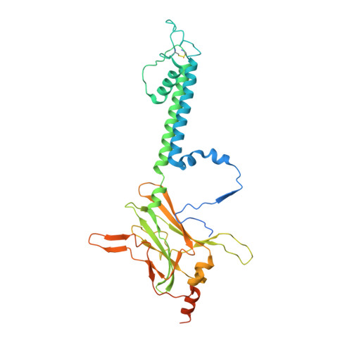
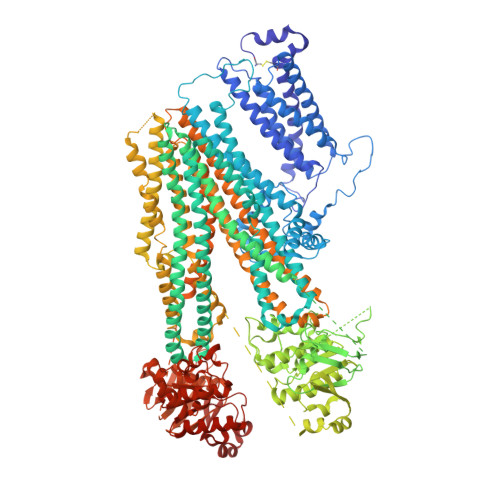
    K ATP channels are metabolic sensors that couple cell energetics to membrane excitability. In pancreatic β-cells, channels formed by SUR1 and Kir6.2 regulate insulin secretion and are the targets of antidiabetic sulfonylureas. Here, we used cryo-EM to elucidate structural basis of channel assembly and gating. The structure, determined in the presence of ATP and the sulfonylurea glibenclamide, at ~6 Å resolution reveals a closed Kir6.2 tetrameric core with four peripheral SUR1s each anchored to a Kir6.2 by its N-terminal transmembrane domain (TMD0). Intricate interactions between TMD0, the loop following TMD0, and Kir6.2 near the proposed PIP 2 binding site, and where ATP density is observed, suggest SUR1 may contribute to ATP and PIP 2 binding to enhance Kir6.2 sensitivity to both. The SUR1-ABC core is found in an unusual inward-facing conformation whereby the two nucleotide binding domains are misaligned along a two-fold symmetry axis, revealing a possible mechanism by which glibenclamide inhibits channel activity.


  • Organizational Affiliation

    Department of Biochemistry and Molecular Biology, Oregon Health and Science University, Portland, Oregon, United States.


Macromolecules
Find similar proteins by:  (by identity cutoff)  |  3D Structure
Entity ID: 1
MoleculeChains Sequence LengthOrganismDetailsImage
ATP-sensitive inward rectifier potassium channel 11
A, C, E, G
390Rattus norvegicusMutation(s): 0 
Gene Names: Kcnj11
Membrane Entity: Yes 
UniProt
Find proteins for P70673 (Rattus norvegicus)
Explore P70673 
Go to UniProtKB:  P70673
Entity Groups  
Sequence Clusters30% Identity50% Identity70% Identity90% Identity95% Identity100% Identity
UniProt GroupP70673
Sequence Annotations
Expand
  • Reference Sequence
Find similar proteins by:  (by identity cutoff)  |  3D Structure
Entity ID: 2
MoleculeChains Sequence LengthOrganismDetailsImage
ATP-binding cassette sub-family C member 8
B, D, F, H
1,590Cricetus cricetusMutation(s): 0 
Gene Names: ABCC8SUR
Membrane Entity: Yes 
UniProt
Find proteins for Q09427 (Cricetus cricetus)
Explore Q09427 
Go to UniProtKB:  Q09427
Entity Groups  
Sequence Clusters30% Identity50% Identity70% Identity90% Identity95% Identity100% Identity
UniProt GroupQ09427
Sequence Annotations
Expand
  • Reference Sequence
Experimental Data & Validation

Experimental Data

  • Method: ELECTRON MICROSCOPY
  • Resolution: 6.30 Å
  • Aggregation State: PARTICLE 
  • Reconstruction Method: SINGLE PARTICLE 
EM Software:
TaskSoftware PackageVersion
RECONSTRUCTIONRELION2.0

Structure Validation

View Full Validation Report


Entry History & Funding Information

Deposition Data


Funding OrganizationLocationGrant Number
National Institutes of Health/National Institute of Diabetes and Digestive and Kidney Disease (NIH/NIDDK)United StatesR01DK066485
National Institutes of Health/National Institute of Diabetes and Digestive and Kidney Disease (NIH/NIDDK)United StatesF31DK105300

Revision History  (Full details and data files)

  • Version 1.0: 2017-01-25
    Type: Initial release
  • Version 1.1: 2017-05-31
    Changes: Structure summary
  • Version 1.2: 2017-09-13
    Changes: Author supporting evidence, Data collection
  • Version 1.3: 2019-12-25
    Changes: Author supporting evidence