5TVM | pdb_00005tvm

Crystal structure of Trypanosoma brucei AdoMetDC/prozyme heterodimer


Experimental Data Snapshot

  • Method: X-RAY DIFFRACTION
  • Resolution: 2.41 Å
  • R-Value Free: 
    0.274 (Depositor), 0.277 (DCC) 
  • R-Value Work: 
    0.229 (Depositor), 0.231 (DCC) 
  • R-Value Observed: 
    0.231 (Depositor) 

Starting Model: experimental
View more details

wwPDB Validation 3D Report Full Report

Validation slider image for 5TVM

This is version 2.1 of the entry. See complete history

Literature

Relief of autoinhibition by conformational switch explains enzyme activation by a catalytically dead paralog.

Volkov, O.A.Kinch, L.N.Ariagno, C.Deng, X.Zhong, S.Grishin, N.V.Tomchick, D.R.Chen, Z.Phillips, M.A.

(2016) Elife 5

  • DOI: https://doi.org/10.7554/eLife.20198
  • Primary Citation Related Structures: 
    5TVF, 5TVM, 5TVO

  • PubMed Abstract: 

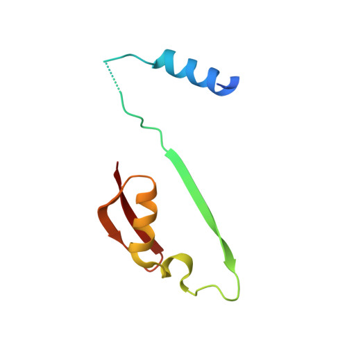
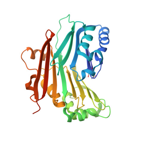
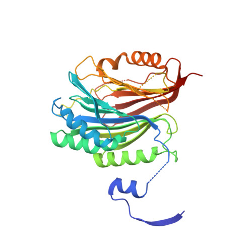
    Catalytically inactive enzyme paralogs occur in many genomes. Some regulate their active counterparts but the structural principles of this regulation remain largely unknown. We report X-ray structures of Trypanosoma brucei S -adenosylmethionine decarboxylase alone and in functional complex with its catalytically dead paralogous partner, prozyme. We show monomeric Tb AdoMetDC is inactive because of autoinhibition by its N-terminal sequence. Heterodimerization with prozyme displaces this sequence from the active site through a complex mechanism involving a cis -to- trans proline isomerization, reorganization of a β-sheet, and insertion of the N-terminal α-helix into the heterodimer interface, leading to enzyme activation. We propose that the evolution of this intricate regulatory mechanism was facilitated by the acquisition of the dimerization domain, a single step that can in principle account for the divergence of regulatory schemes in the AdoMetDC enzyme family. These studies elucidate an allosteric mechanism in an enzyme and a plausible scheme by which such complex cooperativity evolved.


  • Organizational Affiliation
    • Department of Pharmacology, University of Texas Southwestern Medical Center, Dallas, United States.

Macromolecule Content 

  • Total Structure Weight: 157.02 kDa 
  • Atom Count: 10,435 
  • Modeled Residue Count: 1,292 
  • Deposited Residue Count: 1,390 
  • Unique protein chains: 3

Macromolecules

Find similar proteins by:|  3D Structure
Entity ID: 1
MoleculeChains  Sequence LengthOrganismDetailsImage
S-adenosylmethionine decarboxylase beta chain
A, C
85Trypanosoma brucei brucei TREU927Mutation(s): 0 
Gene Names: Tb927.6.4410Tb927.6.4460
EC: 4.1.1.50
UniProt
Find proteins for Q587A7 (Trypanosoma brucei brucei (strain 927/4 GUTat10.1))
Explore Q587A7 
Go to UniProtKB:  Q587A7
Entity Groups
Sequence Clusters30% Identity50% Identity70% Identity90% Identity95% Identity100% Identity
UniProt GroupQ587A7
Sequence Annotations
Expand
Reference Sequence
Find similar proteins by:|  3D Structure
Entity ID: 2
MoleculeChains  Sequence LengthOrganismDetailsImage
S-adenosylmethionine decarboxylase alpha chain
B, D
285Trypanosoma brucei brucei TREU927Mutation(s): 0 
Gene Names: Tb927.6.4410Tb927.6.4460
EC: 4.1.1.50
UniProt
Find proteins for Q587A7 (Trypanosoma brucei brucei (strain 927/4 GUTat10.1))
Explore Q587A7 
Go to UniProtKB:  Q587A7
Entity Groups
Sequence Clusters30% Identity50% Identity70% Identity90% Identity95% Identity100% Identity
UniProt GroupQ587A7
Sequence Annotations
Expand
Reference Sequence
Find similar proteins by:|  3D Structure
Entity ID: 3
MoleculeChains  Sequence LengthOrganismDetailsImage
S-adenosylmethionine decarboxylase proenzyme-like, putative
E, F
325Trypanosoma brucei brucei TREU927Mutation(s): 0 
Gene Names: Tb927.6.4470
EC: 4.1.1.50
UniProt
Find proteins for Q587B3 (Trypanosoma brucei brucei (strain 927/4 GUTat10.1))
Explore Q587B3 
Go to UniProtKB:  Q587B3
Entity Groups
Sequence Clusters30% Identity50% Identity70% Identity90% Identity95% Identity100% Identity
UniProt GroupQ587B3
Sequence Annotations
Expand
Reference Sequence

Experimental Data & Validation

Experimental Data

  • Method: X-RAY DIFFRACTION
  • Resolution: 2.41 Å
  • R-Value Free:  0.274 (Depositor), 0.277 (DCC) 
  • R-Value Work:  0.229 (Depositor), 0.231 (DCC) 
  • R-Value Observed: 0.231 (Depositor) 
Space Group: P 1 21 1
Unit Cell:
Length ( Å )Angle ( ˚ )
a = 81.297α = 90
b = 96.708β = 102.64
c = 99.584γ = 90
Software Package:
Software NamePurpose
PHENIXrefinement
HKL-3000data reduction
HKL-3000data scaling
PHASERphasing

Structure Validation

View Full Validation Report


Entry History 

& Funding Information

Deposition Data


Funding OrganizationLocationGrant Number
National Institutes of Health/National Institute Of Allergy and Infectious Diseases (NIH/NIAID)United States2R37AI034432
National Institutes of Health/National Institute Of Allergy and Infectious Diseases (NIH/NIAID)United StatesR01AI090599

Revision History  (Full details and data files)

  • Version 1.0: 2017-01-11
    Type: Initial release
  • Version 1.1: 2017-09-27
    Changes: Advisory, Author supporting evidence
  • Version 1.2: 2019-12-11
    Changes: Advisory, Author supporting evidence
  • Version 1.3: 2023-10-04
    Changes: Data collection, Database references, Derived calculations, Refinement description
  • Version 2.0: 2023-11-15
    Changes: Atomic model, Data collection, Derived calculations
  • Version 2.1: 2024-11-13
    Changes: Structure summary