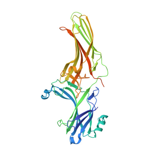
5TV1 | pdb_00005tv1

active arrestin-3 with inositol hexakisphosphate


Experimental Data Snapshot

  • Method: X-RAY DIFFRACTION
  • Resolution: 2.40 Å
  • R-Value Free: 
    0.245 (Depositor), 0.240 (DCC) 
  • R-Value Work: 
    0.197 (Depositor), 0.200 (DCC) 
  • R-Value Observed: 
    0.199 (Depositor) 

Starting Model: experimental
View more details

wwPDB Validation   3D Report Full Report


Ligand Structure Quality Assessment 

Created with Raphaël 2.3.0Worse 01 BetterLigand structure goodness of fit to experimental dataBest fitted IHPClick on this verticalbar to view details

This is version 1.3 of the entry. See complete history


Literature

Structural basis of arrestin-3 activation and signaling.

Chen, Q.Perry, N.A.Vishnivetskiy, S.A.Berndt, S.Gilbert, N.C.Zhuo, Y.Singh, P.K.Tholen, J.Ohi, M.D.Gurevich, E.V.Brautigam, C.A.Klug, C.S.Gurevich, V.V.Iverson, T.M.

(2017) Nat Commun 8: 1427-1427

  • DOI: https://doi.org/10.1038/s41467-017-01218-8
  • Primary Citation of Related Structures:  
    5TV1

  • PubMed Abstract: 

    A unique aspect of arrestin-3 is its ability to support both receptor-dependent and receptor-independent signaling. Here, we show that inositol hexakisphosphate (IP 6 ) is a non-receptor activator of arrestin-3 and report the structure of IP 6 -activated arrestin-3 at 2.4-Å resolution. IP 6 -activated arrestin-3 exhibits an inter-domain twist and a displaced C-tail, hallmarks of active arrestin. IP 6 binds to the arrestin phosphate sensor, and is stabilized by trimerization. Analysis of the trimerization surface, which is also the receptor-binding surface, suggests a feature called the finger loop as a key region of the activation sensor. We show that finger loop helicity and flexibility may underlie coupling to hundreds of diverse receptors and also promote arrestin-3 activation by IP 6 . Importantly, we show that effector-binding sites on arrestins have distinct conformations in the basal and activated states, acting as switch regions. These switch regions may work with the inter-domain twist to initiate and direct arrestin-mediated signaling.


  • Organizational Affiliation

    Department of Pharmacology, Vanderbilt University, Nashville, TN, 37232, USA.


Macromolecules
Find similar proteins by:  (by identity cutoff)  |  3D Structure
Entity ID: 1
MoleculeChains Sequence LengthOrganismDetailsImage
Beta-arrestin-2393Bos taurusMutation(s): 0 
Gene Names: ARRB2
UniProt
Find proteins for P32120 (Bos taurus)
Explore P32120 
Go to UniProtKB:  P32120
Entity Groups  
Sequence Clusters30% Identity50% Identity70% Identity90% Identity95% Identity100% Identity
UniProt GroupP32120
Sequence Annotations
Expand
  • Reference Sequence
Experimental Data & Validation

Experimental Data

  • Method: X-RAY DIFFRACTION
  • Resolution: 2.40 Å
  • R-Value Free:  0.245 (Depositor), 0.240 (DCC) 
  • R-Value Work:  0.197 (Depositor), 0.200 (DCC) 
  • R-Value Observed: 0.199 (Depositor) 
Space Group: P 63
Unit Cell:
Length ( Å )Angle ( ˚ )
a = 97.575α = 90
b = 97.575β = 90
c = 76.938γ = 120
Software Package:
Software NamePurpose
PHENIXrefinement
HKL-2000data scaling
HKL-2000data reduction
PDB_EXTRACTdata extraction
PHASERphasing

Structure Validation

View Full Validation Report


Ligand Structure Quality Assessment 

Created with Raphaël 2.3.0Worse 01 BetterLigand structure goodness of fit to experimental dataBest fitted IHPClick on this verticalbar to view details

Entry History & Funding Information

Deposition Data


Funding OrganizationLocationGrant Number
National Institutes of Health/National Institute of General Medical Sciences (NIH/NIGMS)United StatesGM095633
National Institutes of Health/National Institute of General Medical Sciences (NIH/NIGMS)United StatesGM077561
National Institutes of Health/National Institute of General Medical Sciences (NIH/NIGMS)United StatesGM109955
National Institutes of Health/National Eye Institute (NIH/NEI)United StatesEY011500

Revision History  (Full details and data files)

  • Version 1.0: 2017-11-22
    Type: Initial release
  • Version 1.1: 2019-12-11
    Changes: Author supporting evidence
  • Version 1.2: 2020-10-14
    Changes: Structure summary
  • Version 1.3: 2023-10-04
    Changes: Data collection, Database references, Refinement description