5TUJ | pdb_00005tuj

Ancestral Cationic Amino Acid Solute Binding Protein (AncCDT-1)


Experimental Data Snapshot

  • Method: X-RAY DIFFRACTION
  • Resolution: 3.35 Å
  • R-Value Free: 
    0.338 (Depositor), 0.343 (DCC) 
  • R-Value Work: 
    0.305 (Depositor), 0.312 (DCC) 
  • R-Value Observed: 
    0.307 (Depositor) 

wwPDB Validation 3D Report Full Report

Validation slider image for 5TUJ

This is version 1.2 of the entry. See complete history

Literature

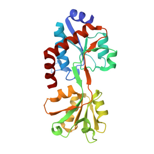
Evolution of cyclohexadienyl dehydratase from an ancestral solute-binding protein.

Clifton, B.E.Kaczmarski, J.A.Carr, P.D.Gerth, M.L.Tokuriki, N.Jackson, C.J.

(2018) Nat Chem Biol 14: 542-547

  • DOI: https://doi.org/10.1038/s41589-018-0043-2
  • Primary Citation Related Structures: 
    5TUJ, 5WJP, 6BQE

  • PubMed Abstract: 

    The emergence of enzymes through the neofunctionalization of noncatalytic proteins is ultimately responsible for the extraordinary range of biological catalysts observed in nature. Although the evolution of some enzymes from binding proteins can be inferred by homology, we have a limited understanding of the nature of the biochemical and biophysical adaptations along these evolutionary trajectories and the sequence in which they occurred. Here we reconstructed and characterized evolutionary intermediate states linking an ancestral solute-binding protein to the extant enzyme cyclohexadienyl dehydratase. We show how the intrinsic reactivity of a desolvated general acid was harnessed by a series of mutations radiating from the active site, which optimized enzyme-substrate complementarity and transition-state stabilization and minimized sampling of noncatalytic conformations. Our work reveals the molecular evolutionary processes that underlie the emergence of enzymes de novo, which are notably mirrored by recent examples of computational enzyme design and directed evolution.


  • Organizational Affiliation
    • Research School of Chemistry, Australian National University, Canberra, ACT, Australia.

Macromolecule Content 

  • Total Structure Weight: 26.29 kDa 
  • Atom Count: 1,738 
  • Modeled Residue Count: 229 
  • Deposited Residue Count: 235 
  • Unique protein chains: 1

Macromolecules

Find similar proteins by:|  3D Structure
Entity ID: 1
MoleculeChains  Sequence LengthOrganismDetailsImage
Ancestral protein CDT-Anc1A [auth C]235unidentifiedMutation(s): 0 

Experimental Data & Validation

Experimental Data

  • Method: X-RAY DIFFRACTION
  • Resolution: 3.35 Å
  • R-Value Free:  0.338 (Depositor), 0.343 (DCC) 
  • R-Value Work:  0.305 (Depositor), 0.312 (DCC) 
  • R-Value Observed: 0.307 (Depositor) 
Space Group: P 32 2 1
Unit Cell:
Length ( Å )Angle ( ˚ )
a = 71.641α = 90
b = 71.641β = 90
c = 124.001γ = 120
Software Package:
Software NamePurpose
PHENIXrefinement
XDSdata reduction
Aimlessdata scaling
PHASERphasing

Structure Validation

View Full Validation Report


Entry History 

Deposition Data

Revision History  (Full details and data files)

  • Version 1.0: 2017-12-06
    Type: Initial release
  • Version 1.1: 2019-01-16
    Changes: Data collection, Database references
  • Version 1.2: 2024-03-06
    Changes: Data collection, Database references