5TTZ | pdb_00005ttz

Crystal structure of Grp94 bound to isoform-selective inhibitor methyl 2-(2-(1-(4-bromobenzyl)-1H-imidazol-2-yl)ethyl)-3-chloro-4,6-dihydroxybenzoate


Experimental Data Snapshot

  • Method: X-RAY DIFFRACTION
  • Resolution: 2.71 Å
  • R-Value Free: 
    0.285 (Depositor), 0.284 (DCC) 
  • R-Value Work: 
    0.199 (Depositor), 0.201 (DCC) 
  • R-Value Observed: 
    0.208 (Depositor) 

Starting Model: experimental
View more details

wwPDB Validation 3D Report Full Report

Validation slider image for 5TTZ

Ligand Structure Quality Assessment 


This is version 1.2 of the entry. See complete history

Literature

Isoform-selective Hsp90 inhibition rescues model of hereditary open-angle glaucoma.

Stothert, A.R.Suntharalingam, A.Tang, X.Crowley, V.M.Mishra, S.J.Webster, J.M.Nordhues, B.A.Huard, D.J.E.Passaglia, C.L.Lieberman, R.L.Blagg, B.S.J.Blair, L.J.Koren, J.Dickey, C.A.

(2017) Sci Rep 7: 17951-17951

  • DOI: https://doi.org/10.1038/s41598-017-18344-4
  • Primary Citation Related Structures: 
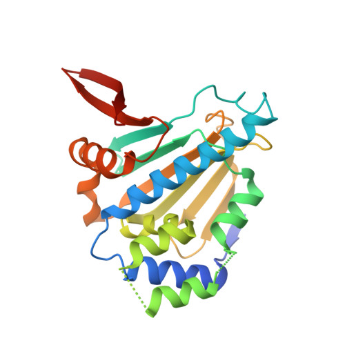
    5TTZ

  • PubMed Abstract: 

    The heat shock protein 90 (Hsp90) family of molecular chaperones regulates protein homeostasis, folding, and degradation. The ER-resident Hsp90 isoform, glucose-regulated protein 94 (Grp94), promotes the aggregation of mutant forms of myocilin, a protein associated with primary open-angle glaucoma. While inhibition of Grp94 promotes the degradation of mutant myocilin in vitro, to date no Grp94-selective inhibitors have been investigated in vivo. Here, a Grp94-selective inhibitor facilitated mutant myocilin degradation and rescued phenotypes in a transgenic mouse model of hereditary primary open-angle glaucoma. Ocular toxicities previously associated with pan-Hsp90 inhibitors were not evident with our Grp94-selective inhibitor, 4-Br-BnIm. Our study suggests that selective inhibition of a distinct Hsp90 family member holds translational promise for ocular and other diseases associated with cell stress and protein misfolding.


  • Organizational Affiliation
    • Department of Molecular Medicine and Byrd Alzheimer's Research Institute, University of South Florida, Tampa, FL, 33613, USA.

Macromolecule Content 

  • Total Structure Weight: 54.11 kDa 
  • Atom Count: 3,454 
  • Modeled Residue Count: 428 
  • Deposited Residue Count: 466 
  • Unique protein chains: 1

Macromolecules

Find similar proteins by:|  3D Structure
Entity ID: 1
MoleculeChains  Sequence LengthOrganismDetailsImage
Endoplasmin
A, B
233Canis lupus familiarisMutation(s): 0 
Gene Names: HSP90B1GRP94TRA1
EC: 3.6.4
UniProt
Find proteins for P41148 (Canis lupus familiaris)
Explore P41148 
Go to UniProtKB:  P41148
Entity Groups
Sequence Clusters30% Identity50% Identity70% Identity90% Identity95% Identity100% Identity
UniProt GroupP41148
Sequence Annotations
Expand
Reference Sequence

Small Molecules

Ligands 5 Unique
IDChains Name / Formula / InChI Key2D Diagram3D Interactions
7KY

Query on 7KY


Download:Ideal Coordinates CCD File
C [auth A],
E [auth B]
methyl 2-(2-{1-[(4-bromophenyl)methyl]-1H-imidazol-2-yl}ethyl)-3-chloro-4,6-dihydroxybenzoate
C20 H18 Br Cl N2 O4
FHDUIAYTJZAXNW-UHFFFAOYSA-N
PE4

Query on PE4


Download:Ideal Coordinates CCD File
H [auth B]2-{2-[2-(2-{2-[2-(2-ETHOXY-ETHOXY)-ETHOXY]-ETHOXY}-ETHOXY)-ETHOXY]-ETHOXY}-ETHANOL
C16 H34 O8
PJWQOENWHPEPKI-UHFFFAOYSA-N
PG4

Query on PG4


Download:Ideal Coordinates CCD File
F [auth B]TETRAETHYLENE GLYCOL
C8 H18 O5
UWHCKJMYHZGTIT-UHFFFAOYSA-N
PGE

Query on PGE


Download:Ideal Coordinates CCD File
G [auth B]TRIETHYLENE GLYCOL
C6 H14 O4
ZIBGPFATKBEMQZ-UHFFFAOYSA-N
PEG

Query on PEG


Download:Ideal Coordinates CCD File
D [auth A]DI(HYDROXYETHYL)ETHER
C4 H10 O3
MTHSVFCYNBDYFN-UHFFFAOYSA-N

Experimental Data & Validation

Experimental Data

  • Method: X-RAY DIFFRACTION
  • Resolution: 2.71 Å
  • R-Value Free:  0.285 (Depositor), 0.284 (DCC) 
  • R-Value Work:  0.199 (Depositor), 0.201 (DCC) 
  • R-Value Observed: 0.208 (Depositor) 
Space Group: P 21 21 21
Unit Cell:
Length ( Å )Angle ( ˚ )
a = 65.237α = 90
b = 84.42β = 90
c = 95.473γ = 90
Software Package:
Software NamePurpose
PHENIXrefinement
XDSdata reduction
XSCALEdata scaling
PHASERphasing

Structure Validation

View Full Validation Report


Ligand Structure Quality Assessment 


Entry History 

& Funding Information

Deposition Data


Funding OrganizationLocationGrant Number
National Institutes of Health/National Eye Institute (NIH/NEI)United StatesR01EY021205

Revision History  (Full details and data files)

  • Version 1.0: 2018-01-10
    Type: Initial release
  • Version 1.1: 2019-12-11
    Changes: Author supporting evidence
  • Version 1.2: 2023-10-04
    Changes: Advisory, Data collection, Database references, Refinement description