


Novel Enhancer Binding Site Found In Bacteria And Eukaryota But Not In Archea.
Dimova, M., Devedjiev, Y.D.To be published.
Experimental Data Snapshot
Starting Model: experimental
View more details
Entity ID: 1 | |||||
|---|---|---|---|---|---|
| Molecule | Chains | Sequence Length | Organism | Details | Image |
| Glyceraldehyde-3-phosphate dehydrogenase | A [auth P], B [auth R], C [auth S] | 333 | Sus scrofa | Mutation(s): 0 EC: 1.2.1.12 (PDB Primary Data), 2.6.99 (PDB Primary Data) | 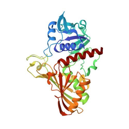 |
UniProt | |||||
Entity Groups | |||||
| Sequence Clusters | 30% Identity50% Identity70% Identity90% Identity95% Identity100% Identity | ||||
| UniProt Group | P00355 | ||||
Sequence AnnotationsExpand | |||||
Reference Sequence | |||||
| Ligands 4 Unique | |||||
|---|---|---|---|---|---|
| ID | Chains | Name / Formula / InChI Key | 2D Diagram | 3D Interactions | |
| NAD Download:Ideal Coordinates CCD File | F [auth P], O [auth R], X [auth S] | NICOTINAMIDE-ADENINE-DINUCLEOTIDE C21 H27 N7 O14 P2 BAWFJGJZGIEFAR-NNYOXOHSSA-N |  | ||
| PHN Download:Ideal Coordinates CCD File | K [auth R], L [auth R], T [auth S], U [auth S] | 1,10-PHENANTHROLINE C12 H8 N2 DGEZNRSVGBDHLK-UHFFFAOYSA-N |  | ||
| SO4 Download:Ideal Coordinates CCD File | D [auth P] E [auth P] G [auth P] H [auth P] I [auth P] | SULFATE ION O4 S QAOWNCQODCNURD-UHFFFAOYSA-L |  | ||
| GOL Download:Ideal Coordinates CCD File | AA [auth S] BA [auth S] J [auth P] R S [auth R] | GLYCEROL C3 H8 O3 PEDCQBHIVMGVHV-UHFFFAOYSA-N |  | ||
| Length ( Å ) | Angle ( ˚ ) |
|---|---|
| a = 86.48 | α = 90 |
| b = 133.52 | β = 90 |
| c = 210.32 | γ = 90 |
| Software Name | Purpose |
|---|---|
| REFMAC | refinement |
| SCALEPACK | data scaling |
| AMoRE | phasing |
| DENZO | data reduction |


