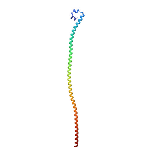
5TO6

Structure of the TPR oligomerization domain


Experimental Data Snapshot

  • Method: X-RAY DIFFRACTION
  • Resolution: 2.70 Å
  • R-Value Free: 0.288 
  • R-Value Work: 0.237 
  • R-Value Observed: 0.239 

Starting Model: experimental
View more details

wwPDB Validation   3D Report Full Report


This is version 1.3 of the entry. See complete history


Literature

Structural Basis of TPR-Mediated Oligomerization and Activation of Oncogenic Fusion Kinases.

Pal, K.Bandyopadhyay, A.Zhou, X.E.Xu, Q.Marciano, D.P.Brunzelle, J.S.Yerrum, S.Griffin, P.R.Vande Woude, G.Melcher, K.Xu, H.E.

(2017) Structure 25: 867-877.e3

  • DOI: https://doi.org/10.1016/j.str.2017.04.015
  • Primary Citation of Related Structures:  
    5TO5, 5TO6, 5TO7, 5TVB

  • PubMed Abstract: 

    The nuclear pore complex subunit TPR is found in at least five different oncogenic fusion kinases, including TPR-MET, yet how TPR fusions promote activation of kinases and their oncogenic activities remains poorly understood. Here we report the crystal structure of TPR(2-142), the MET fusion partner of oncogenic TPR-MET. TPR(2-142) contains a continuous 124-residue α helix that forms an antiparallel tetramer from two leucine zipper-containing parallel coiled coils. Remarkably, single mutations cause strikingly different conformations of the coiled coil, indicating its highly dynamic nature. We further show that fusion of TPR(2-142) to the MET intracellular domain strongly and selectively stabilizes the αG helix of the MET kinase domain, and mutations of only the TPR leucine zipper residues at the junction to MET, but not other leucine zipper residues, abolish kinase activation. Together, these results provide critical insight into the TPR structure and its ability to induce dimerization and activation of fusion kinases.


  • Organizational Affiliation

    Center for Cancer and Cell Biology, Van Andel Research Institute, Grand Rapids, MI 49503, USA.


Macromolecules
Find similar proteins by:  (by identity cutoff)  |  3D Structure
Entity ID: 1
MoleculeChains Sequence LengthOrganismDetailsImage
Nucleoprotein TPR
A, B, C, D
141Homo sapiensMutation(s): 0 
Gene Names: TPR
UniProt & NIH Common Fund Data Resources
Find proteins for P12270 (Homo sapiens)
Explore P12270 
Go to UniProtKB:  P12270
PHAROS:  P12270
GTEx:  ENSG00000047410 
Entity Groups  
Sequence Clusters30% Identity50% Identity70% Identity90% Identity95% Identity100% Identity
UniProt GroupP12270
Sequence Annotations
Expand
  • Reference Sequence
Experimental Data & Validation

Experimental Data

  • Method: X-RAY DIFFRACTION
  • Resolution: 2.70 Å
  • R-Value Free: 0.288 
  • R-Value Work: 0.237 
  • R-Value Observed: 0.239 
  • Space Group: P 1 21 1
Unit Cell:
Length ( Å )Angle ( ˚ )
a = 76.79α = 90
b = 34.86β = 91.85
c = 113.47γ = 90
Software Package:
Software NamePurpose
PDB_EXTRACTdata extraction
HKL-2000data reduction
HKL-2000data scaling
MOLREPphasing
PHENIXrefinement

Structure Validation

View Full Validation Report


Entry History 

Deposition Data

Revision History  (Full details and data files)

  • Version 1.0: 2017-10-18
    Type: Initial release
  • Version 1.1: 2019-05-01
    Changes: Data collection, Database references
  • Version 1.2: 2023-10-04
    Changes: Data collection, Database references, Refinement description
  • Version 1.3: 2024-11-06
    Changes: Structure summary