5TKF | pdb_00005tkf

Neurospora crassa polysaccharide monooxygenase 2 high mannosylation


Experimental Data Snapshot

  • Method: X-RAY DIFFRACTION
  • Resolution: 2.10 Å
  • R-Value Free: 
    0.177 (Depositor), 0.182 (DCC) 
  • R-Value Work: 
    0.142 (Depositor), 0.146 (DCC) 
  • R-Value Observed: 
    0.143 (Depositor) 

Starting Model: experimental
View more details

wwPDB Validation 3D Report Full Report

Validation slider image for 5TKF

Ligand Structure Quality Assessment 


This is version 2.2 of the entry. See complete history

Literature

Crystallization of a fungal lytic polysaccharide monooxygenase expressed from glycoengineered Pichia pastoris for X-ray and neutron diffraction.

O'Dell, W.B.Swartz, P.D.Weiss, K.L.Meilleur, F.

(2017) Acta Crystallogr F Struct Biol Commun 73: 70-78

  • DOI: https://doi.org/10.1107/S2053230X16020318
  • Primary Citation Related Structures: 
    5TKF

  • PubMed Abstract: 

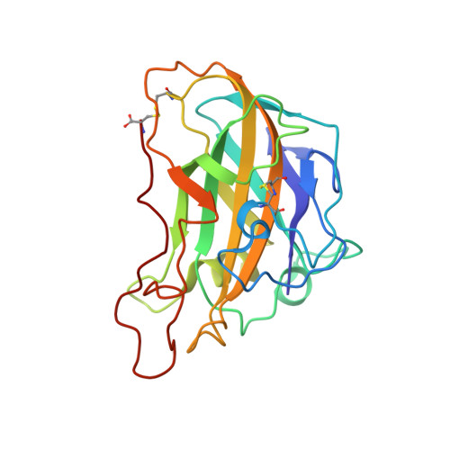
    Lytic polysaccharide monooxygenases (LPMOs) are carbohydrate-disrupting enzymes secreted by bacteria and fungi that break glycosidic bonds via an oxidative mechanism. Fungal LPMOs typically act on cellulose and can enhance the efficiency of cellulose-hydrolyzing enzymes that release soluble sugars for bioethanol production or other industrial uses. The enzyme PMO-2 from Neurospora crassa (NcPMO-2) was heterologously expressed in Pichia pastoris to facilitate crystallographic studies of the fungal LPMO mechanism. Diffraction resolution and crystal morphology were improved by expressing NcPMO-2 from a glycoengineered strain of P. pastoris and by the use of crystal seeding methods, respectively. These improvements resulted in high-resolution (1.20 Å) X-ray diffraction data collection at 100 K and the production of a large NcPMO-2 crystal suitable for room-temperature neutron diffraction data collection to 2.12 Å resolution.


  • Organizational Affiliation
    • Department of Molecular and Structural Biochemistry, North Carolina State University, Campus Box 7622, Raleigh, NC 27695, USA.

Macromolecule Content 

  • Total Structure Weight: 96.14 kDa 
  • Atom Count: 7,858 
  • Modeled Residue Count: 892 
  • Deposited Residue Count: 892 
  • Unique protein chains: 1

Macromolecules

Find similar proteins by:|  3D Structure
Entity ID: 1
MoleculeChains  Sequence LengthOrganismDetailsImage
Lytic polysaccharide monooxygenase
A, B, C, D
223Neurospora crassaMutation(s): 0 
Gene Names: G15G9.090GE21DRAFT_7469
EC: 1.14.99.56
UniProt
Find proteins for Q1K8B6 (Neurospora crassa (strain ATCC 24698 / 74-OR23-1A / CBS 708.71 / DSM 1257 / FGSC 987))
Explore Q1K8B6 
Go to UniProtKB:  Q1K8B6
Entity Groups
Sequence Clusters30% Identity50% Identity70% Identity90% Identity95% Identity100% Identity
UniProt GroupQ1K8B6
Glycosylation
Glycosylation Sites: 1
Sequence Annotations
Expand
Reference Sequence

Oligosaccharides

Help  
Entity ID: 2
MoleculeChains Length2D Diagram GlycosylationD Interactions
alpha-D-mannopyranose-(1-2)-alpha-D-mannopyranose-(1-3)-[alpha-D-mannopyranose-(1-3)-alpha-D-mannopyranose-(1-6)]alpha-D-mannopyranose-(1-4)-2-acetamido-2-deoxy-beta-D-glucopyranose-(1-4)-2-acetamido-2-deoxy-beta-D-glucopyranose
E
7N-Glycosylation
Glycosylation Resources
GlyTouCan: G84676UY
GlyCosmos: G84676UY
GlyGen: G84676UY
Entity ID: 3
MoleculeChains Length2D Diagram GlycosylationD Interactions
2-acetamido-2-deoxy-beta-D-glucopyranose-(1-4)-2-acetamido-2-deoxy-beta-D-glucopyranose
F
2N-Glycosylation
Glycosylation Resources
GlyTouCan: G42666HT
GlyCosmos: G42666HT
GlyGen: G42666HT
Entity ID: 4
MoleculeChains Length2D Diagram GlycosylationD Interactions
alpha-D-mannopyranose-(1-3)-alpha-D-mannopyranose-(1-4)-2-acetamido-2-deoxy-beta-D-glucopyranose-(1-4)-2-acetamido-2-deoxy-beta-D-glucopyranose
G
4N-Glycosylation
Glycosylation Resources
GlyTouCan: G48068RF
GlyCosmos: G48068RF
GlyGen: G48068RF

Small Molecules

Ligands 3 Unique
IDChains Name / Formula / InChI Key2D Diagram3D Interactions
NAG

Query on NAG


Download:Ideal Coordinates CCD File
L [auth D]2-acetamido-2-deoxy-beta-D-glucopyranose
C8 H15 N O6
OVRNDRQMDRJTHS-FMDGEEDCSA-N
CU

Query on CU


Download:Ideal Coordinates CCD File
H [auth A],
J [auth B],
K [auth C],
M [auth D]
COPPER (II) ION
Cu
JPVYNHNXODAKFH-UHFFFAOYSA-N
OXY

Query on OXY


Download:Ideal Coordinates CCD File
I [auth B],
N [auth D]
OXYGEN MOLECULE
O2
MYMOFIZGZYHOMD-UHFFFAOYSA-N

Experimental Data & Validation

Experimental Data

  • Method: X-RAY DIFFRACTION
  • Resolution: 2.10 Å
  • R-Value Free:  0.177 (Depositor), 0.182 (DCC) 
  • R-Value Work:  0.142 (Depositor), 0.146 (DCC) 
  • R-Value Observed: 0.143 (Depositor) 
Space Group: P 1
Unit Cell:
Length ( Å )Angle ( ˚ )
a = 43.3α = 97.57
b = 67.04β = 97.61
c = 84.15γ = 97.43
Software Package:
Software NamePurpose
PHENIXrefinement
MOSFLMdata reduction
Aimlessdata scaling
PHASERphasing
PDB_EXTRACTdata extraction

Structure Validation

View Full Validation Report


Ligand Structure Quality Assessment 


Entry History 

& Funding Information

Deposition Data


Funding OrganizationLocationGrant Number
National Science Foundation (NSF, United States)United States1069091

Revision History  (Full details and data files)

  • Version 1.0: 2017-05-17
    Type: Initial release
  • Version 1.1: 2017-09-20
    Changes: Author supporting evidence
  • Version 1.2: 2019-11-27
    Changes: Author supporting evidence
  • Version 2.0: 2020-07-29
    Type: Remediation
    Reason: Carbohydrate remediation
    Changes: Atomic model, Data collection, Derived calculations, Structure summary
  • Version 2.1: 2023-10-04
    Changes: Data collection, Database references, Derived calculations, Refinement description, Structure summary
  • Version 2.2: 2024-11-20
    Changes: Structure summary