5THP

Rhodocetin in complex with the integrin alpha2-A domain


Experimental Data Snapshot

  • Method: X-RAY DIFFRACTION
  • Resolution: 3.01 Å
  • R-Value Free: 0.270 
  • R-Value Work: 0.221 
  • R-Value Observed: 0.224 

Starting Model: experimental
View more details

wwPDB Validation   3D Report Full Report


This is version 1.1 of the entry. See complete history


Literature

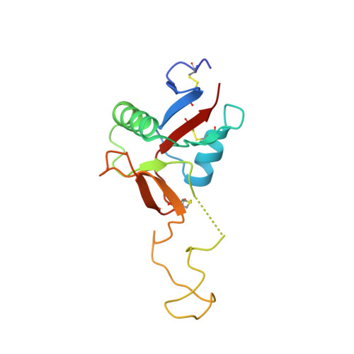
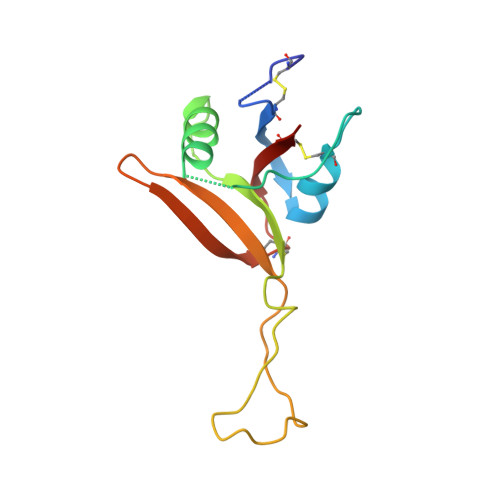
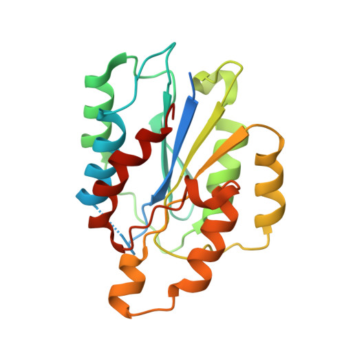
Dramatic and concerted conformational changes enable rhodocetin to block alpha 2 beta 1 integrin selectively.

Eble, J.A.McDougall, M.Orriss, G.L.Niland, S.Johanningmeier, B.Pohlentz, G.Meier, M.Karrasch, S.Estevao-Costa, M.I.Martins Lima, A.Stetefeld, J.

(2017) PLoS Biol 15: e2001492-e2001492

  • DOI: https://doi.org/10.1371/journal.pbio.2001492
  • Primary Citation of Related Structures:  
    5THP

  • PubMed Abstract: 

    The collagen binding integrin α2β1 plays a crucial role in hemostasis, fibrosis, and cancer progression amongst others. It is specifically inhibited by rhodocetin (RC), a C-type lectin-related protein (CLRP) found in Malayan pit viper (Calloselasma rhodostoma) venom. The structure of RC alone reveals a heterotetramer arranged as an αβ and γδ subunit in a cruciform shape. RC specifically binds to the collagen binding A-domain of the integrin α2 subunit, thereby blocking collagen-induced platelet aggregation. However, until now, the molecular basis for this interaction has remained unclear. Here, we present the molecular structure of the RCγδ-α2A complex solved to 3.0 Å resolution. Our findings show that RC undergoes a dramatic structural reorganization upon binding to α2β1 integrin. Besides the release of the nonbinding RCαβ tandem, the RCγ subunit interacts with loop 2 of the α2A domain as result of a dramatic conformational change. The RCδ subunit contacts the integrin α2A domain in the "closed" conformation through its helix C. Combined with epitope-mapped antibodies, conformationally locked α2A domain mutants, point mutations within the α2A loop 2, and chemical modifications of the purified toxin protein, this molecular structure of RCγδ-α2A complex explains the inhibitory mechanism and specificity of RC for α2β1 integrin.


  • Organizational Affiliation

    Institute of Physiological Chemistry and Pathobiochemistry, University of Münster, Münster, Germany.


Macromolecules
Find similar proteins by:  (by identity cutoff)  |  3D Structure
Entity ID: 1
MoleculeChains Sequence LengthOrganismDetailsImage
Snaclec rhodocetin subunit gamma
A, D, G, J, M
A, D, G, J, M, P
135Calloselasma rhodostomaMutation(s): 0 
UniProt
Find proteins for D2YW39 (Calloselasma rhodostoma)
Explore D2YW39 
Go to UniProtKB:  D2YW39
Entity Groups  
Sequence Clusters30% Identity50% Identity70% Identity90% Identity95% Identity100% Identity
UniProt GroupD2YW39
Sequence Annotations
Expand
  • Reference Sequence
Find similar proteins by:  (by identity cutoff)  |  3D Structure
Entity ID: 2
MoleculeChains Sequence LengthOrganismDetailsImage
Snaclec rhodocetin subunit delta
B, E, H, K, N
B, E, H, K, N, Q
124Calloselasma rhodostomaMutation(s): 0 
UniProt
Find proteins for D2YW40 (Calloselasma rhodostoma)
Explore D2YW40 
Go to UniProtKB:  D2YW40
Entity Groups  
Sequence Clusters30% Identity50% Identity70% Identity90% Identity95% Identity100% Identity
UniProt GroupD2YW40
Sequence Annotations
Expand
  • Reference Sequence
Find similar proteins by:  (by identity cutoff)  |  3D Structure
Entity ID: 3
MoleculeChains Sequence LengthOrganismDetailsImage
Integrin alpha-2
C, F, I, L, O
C, F, I, L, O, R
217Homo sapiensMutation(s): 0 
Gene Names: ITGA2CD49B
UniProt & NIH Common Fund Data Resources
Find proteins for P17301 (Homo sapiens)
Explore P17301 
Go to UniProtKB:  P17301
GTEx:  ENSG00000164171 
Entity Groups  
Sequence Clusters30% Identity50% Identity70% Identity90% Identity95% Identity100% Identity
UniProt GroupP17301
Sequence Annotations
Expand
  • Reference Sequence
Small Molecules
Ligands 5 Unique
IDChains Name / Formula / InChI Key2D Diagram3D Interactions
SO4
Query on SO4

Download Ideal Coordinates CCD File 
CB [auth F]
DC [auth L]
DD [auth R]
EA [auth C]
MC [auth O]
CB [auth F],
DC [auth L],
DD [auth R],
EA [auth C],
MC [auth O],
OA [auth D],
RC [auth O],
SB [auth I],
WA [auth E],
XA [auth F]
SULFATE ION
O4 S
QAOWNCQODCNURD-UHFFFAOYSA-L
GOL
Query on GOL

Download Ideal Coordinates CCD File 
FA [auth C]
GB [auth G]
HB [auth G]
LB [auth H]
U [auth A]
FA [auth C],
GB [auth G],
HB [auth G],
LB [auth H],
U [auth A],
VC [auth P],
WB [auth J],
XB [auth J]
GLYCEROL
C3 H8 O3
PEDCQBHIVMGVHV-UHFFFAOYSA-N
CL
Query on CL

Download Ideal Coordinates CCD File 
AA [auth C]
BA [auth C]
BB [auth F]
BC [auth L]
CA [auth C]
AA [auth C],
BA [auth C],
BB [auth F],
BC [auth L],
CA [auth C],
CC [auth L],
CD [auth R],
DA [auth C],
EB [auth G],
FB [auth G],
FC [auth M],
GC [auth M],
IC [auth N],
JA [auth D],
JB [auth H],
JC [auth N],
KA [auth D],
KB [auth H],
KC [auth N],
LA [auth D],
MA [auth D],
NA [auth D],
PB [auth I],
QB [auth I],
QC [auth O],
RB [auth I],
T [auth A],
TA [auth E],
TC [auth P],
UA [auth E],
UC [auth P],
VA [auth E],
VB [auth J],
YC [auth Q],
ZC [auth Q]
CHLORIDE ION
Cl
VEXZGXHMUGYJMC-UHFFFAOYSA-M
MG
Query on MG

Download Ideal Coordinates CCD File 
AB [auth F]
AC [auth L]
BD [auth R]
OB [auth I]
PC [auth O]
AB [auth F],
AC [auth L],
BD [auth R],
OB [auth I],
PC [auth O],
Z [auth C]
MAGNESIUM ION
Mg
JLVVSXFLKOJNIY-UHFFFAOYSA-N
NA
Query on NA

Download Ideal Coordinates CCD File 
AD [auth R]
DB [auth G]
EC [auth M]
GA [auth C]
HA [auth D]
AD [auth R],
DB [auth G],
EC [auth M],
GA [auth C],
HA [auth D],
HC [auth N],
IA [auth D],
IB [auth H],
LC [auth O],
MB [auth I],
NB [auth I],
NC [auth O],
OC [auth O],
PA [auth D],
QA [auth E],
RA [auth E],
S [auth A],
SA [auth E],
SC [auth P],
TB [auth J],
UB [auth J],
V [auth B],
W [auth C],
WC [auth Q],
X [auth C],
XC [auth Q],
Y [auth C],
YA [auth F],
YB [auth L],
ZA [auth F],
ZB [auth L]
SODIUM ION
Na
FKNQFGJONOIPTF-UHFFFAOYSA-N
Experimental Data & Validation

Experimental Data

  • Method: X-RAY DIFFRACTION
  • Resolution: 3.01 Å
  • R-Value Free: 0.270 
  • R-Value Work: 0.221 
  • R-Value Observed: 0.224 
  • Space Group: P 41
Unit Cell:
Length ( Å )Angle ( ˚ )
a = 130.763α = 90
b = 130.763β = 90
c = 251.351γ = 90
Software Package:
Software NamePurpose
PHENIXrefinement
XDSdata reduction
Aimlessdata scaling
PHASERphasing

Structure Validation

View Full Validation Report


Entry History & Funding Information

Deposition Data


Funding OrganizationLocationGrant Number
Heart and Stroke Foundation Canada, Grant-in-Aid programCanadaG-14-0006025

Revision History  (Full details and data files)

  • Version 1.0: 2017-08-02
    Type: Initial release
  • Version 1.1: 2023-10-04
    Changes: Data collection, Database references, Derived calculations, Refinement description