5T89

Crystal structure of VEGF-A in complex with VEGFR-1 domains D1-6


Experimental Data Snapshot

  • Method: X-RAY DIFFRACTION
  • Resolution: 4.00 Å
  • R-Value Free: 0.315 
  • R-Value Work: 0.285 
  • R-Value Observed: 0.286 

wwPDB Validation   3D Report Full Report


Ligand Structure Quality Assessment 


This is version 2.0 of the entry. See complete history


Literature

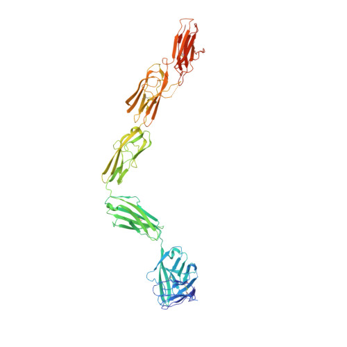
Structure of the Full-length VEGFR-1 Extracellular Domain in Complex with VEGF-A.

Markovic-Mueller, S.Stuttfeld, E.Asthana, M.Weinert, T.Bliven, S.Goldie, K.N.Kisko, K.Capitani, G.Ballmer-Hofer, K.

(2017) Structure 25: 341-352

  • DOI: https://doi.org/10.1016/j.str.2016.12.012
  • Primary Citation of Related Structures:  
    5T89

  • PubMed Abstract: 

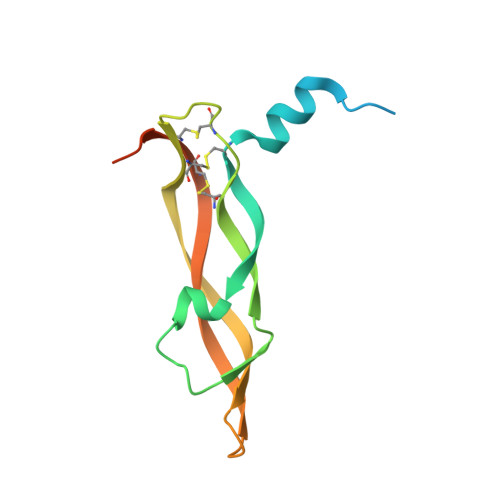
    Vascular endothelial growth factors (VEGFs) regulate blood and lymph vessel development upon activation of three receptor tyrosine kinases: VEGFR-1, -2, and -3. Partial structures of VEGFR/VEGF complexes based on single-particle electron microscopy, small-angle X-ray scattering, and X-ray crystallography revealed the location of VEGF binding and domain arrangement of individual receptor subdomains. Here, we describe the structure of the full-length VEGFR-1 extracellular domain in complex with VEGF-A at 4 Å resolution. We combined X-ray crystallography, single-particle electron microscopy, and molecular modeling for structure determination and validation. The structure reveals the molecular details of ligand-induced receptor dimerization, in particular of homotypic receptor interactions in immunoglobulin homology domains 4, 5, and 7. Functional analyses of ligand binding and receptor activation confirm the relevance of these homotypic contacts and identify them as potential therapeutic sites to allosterically inhibit VEGFR-1 activity.


  • Organizational Affiliation

    Paul Scherrer Institute, Laboratory of Biomolecular Research, 5232 Villigen, Switzerland.


Macromolecules
Find similar proteins by:  (by identity cutoff)  |  3D Structure
Entity ID: 1
MoleculeChains Sequence LengthOrganismDetailsImage
Vascular endothelial growth factor AA [auth V],
B [auth W]
131Homo sapiensMutation(s): 0 
Gene Names: VEGFAVEGF
UniProt & NIH Common Fund Data Resources
Find proteins for P15692 (Homo sapiens)
Explore P15692 
Go to UniProtKB:  P15692
PHAROS:  P15692
GTEx:  ENSG00000112715 
Entity Groups  
Sequence Clusters30% Identity50% Identity70% Identity90% Identity95% Identity100% Identity
UniProt GroupP15692
Sequence Annotations
Expand
  • Reference Sequence
Find similar proteins by:  (by identity cutoff)  |  3D Structure
Entity ID: 2
MoleculeChains Sequence LengthOrganismDetailsImage
Vascular endothelial growth factor receptor 1C [auth X],
D [auth Y]
646Homo sapiensMutation(s): 0 
Gene Names: FLT1FLTFRTVEGFR1
EC: 2.7.10.1
UniProt & NIH Common Fund Data Resources
Find proteins for P17948 (Homo sapiens)
Explore P17948 
Go to UniProtKB:  P17948
PHAROS:  P17948
GTEx:  ENSG00000102755 
Entity Groups  
Sequence Clusters30% Identity50% Identity70% Identity90% Identity95% Identity100% Identity
UniProt GroupP17948
Sequence Annotations
Expand
  • Reference Sequence
Oligosaccharides

Help

Entity ID: 3
MoleculeChains Length2D Diagram Glycosylation3D Interactions
2-acetamido-2-deoxy-beta-D-glucopyranose-(1-4)-2-acetamido-2-deoxy-beta-D-glucopyranoseE [auth A],
F [auth B]
2N-Glycosylation
Glycosylation Resources
GlyTouCan:  G42666HT
GlyCosmos:  G42666HT
GlyGen:  G42666HT
Small Molecules
Ligands 1 Unique
IDChains Name / Formula / InChI Key2D Diagram3D Interactions
NAG
Query on NAG

Download Ideal Coordinates CCD File 
G [auth V]
H [auth W]
I [auth X]
J [auth X]
K [auth X]
G [auth V],
H [auth W],
I [auth X],
J [auth X],
K [auth X],
L [auth X],
M [auth X],
N [auth X],
O [auth X],
P [auth X],
Q [auth Y],
R [auth Y],
S [auth Y],
T [auth Y],
U [auth Y],
V [auth Y],
W [auth Y],
X [auth Y],
Y
2-acetamido-2-deoxy-beta-D-glucopyranose
C8 H15 N O6
OVRNDRQMDRJTHS-FMDGEEDCSA-N
Experimental Data & Validation

Experimental Data

  • Method: X-RAY DIFFRACTION
  • Resolution: 4.00 Å
  • R-Value Free: 0.315 
  • R-Value Work: 0.285 
  • R-Value Observed: 0.286 
  • Space Group: C 1 2 1
Unit Cell:
Length ( Å )Angle ( ˚ )
a = 166.71α = 90
b = 123.09β = 109.56
c = 167.37γ = 90
Software Package:
Software NamePurpose
PHENIXrefinement
XSCALEdata scaling
PDB_EXTRACTdata extraction
XDSdata reduction
PHASERphasing

Structure Validation

View Full Validation Report


Ligand Structure Quality Assessment 


Entry History & Funding Information

Deposition Data


Funding OrganizationLocationGrant Number
Swiss National Science FoundationSwitzerland31003A-130463
OncosuisseSwitzerlandOC2 01200-08-2007

Revision History  (Full details and data files)

  • Version 1.0: 2017-02-01
    Type: Initial release
  • Version 1.1: 2017-02-15
    Changes: Database references
  • Version 1.2: 2017-09-13
    Changes: Advisory, Author supporting evidence, Data collection
  • Version 1.3: 2017-10-11
    Changes: Data collection
  • Version 2.0: 2020-07-29
    Type: Remediation
    Reason: Carbohydrate remediation
    Changes: Advisory, Atomic model, Data collection, Derived calculations, Structure summary