5T7R | pdb_00005t7r

A6-A11 trans-dicarba human insulin


Experimental Data Snapshot

  • Method: X-RAY DIFFRACTION
  • Resolution: 1.55 Å
  • R-Value Free: 
    0.217 (Depositor), 0.221 (DCC) 
  • R-Value Work: 
    0.194 (Depositor), 0.195 (DCC) 
  • R-Value Observed: 
    0.195 (Depositor) 

wwPDB Validation 3D Report Full Report

Validation slider image for 5T7R

This is version 1.3 of the entry. See complete history

Literature

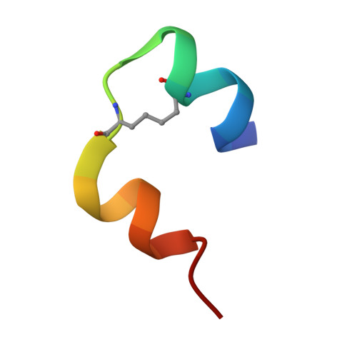
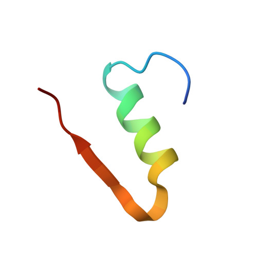
Insulin in motion: The A6-A11 disulfide bond allosterically modulates structural transitions required for insulin activity.

van Lierop, B.Ong, S.C.Belgi, A.Delaine, C.Andrikopoulos, S.Haworth, N.L.Menting, J.G.Lawrence, M.C.Robinson, A.J.Forbes, B.E.

(2017) Sci Rep 7: 17239-17239

  • DOI: https://doi.org/10.1038/s41598-017-16876-3
  • Primary Citation Related Structures: 
    5T7R

  • PubMed Abstract: 

    The structural transitions required for insulin to activate its receptor and initiate regulation of glucose homeostasis are only partly understood. Here, using ring-closing metathesis, we substitute the A6-A11 disulfide bond of insulin with a rigid, non-reducible dicarba linkage, yielding two distinct stereo-isomers (cis and trans). Remarkably, only the cis isomer displays full insulin potency, rapidly lowering blood glucose in mice (even under insulin-resistant conditions). It also posseses reduced mitogenic activity in vitro. Further biophysical, crystallographic and molecular-dynamics analyses reveal that the A6-A11 bond configuration directly affects the conformational flexibility of insulin A-chain N-terminal helix, dictating insulin's ability to engage its receptor. We reveal that in native insulin, contraction of the C α -C α distance of the flexible A6-A11 cystine allows the A-chain N-terminal helix to unwind to a conformation that allows receptor engagement. This motion is also permitted in the cis isomer, with its shorter C α -C α distance, but prevented in the extended trans analogue. These findings thus illuminate for the first time the allosteric role of the A6-A11 bond in mediating the transition of the hormone to an active conformation, significantly advancing our understanding of insulin action and opening up new avenues for the design of improved therapeutic analogues.


  • Organizational Affiliation
    • School of Chemistry, Monash University, Clayton, Victoria, 3800, Australia.

Macromolecule Content 

  • Total Structure Weight: 11.56 kDa 
  • Atom Count: 916 
  • Modeled Residue Count: 100 
  • Deposited Residue Count: 102 
  • Unique protein chains: 2

Macromolecules

Find similar proteins by:  Sequence   |   3D Structure  
Entity ID: 1
MoleculeChains  Sequence LengthOrganismDetailsImage
Insulin A chain
A, C
21Homo sapiensMutation(s): 2 
UniProt & NIH Common Fund Data Resources
Find proteins for P01308 (Homo sapiens)
Explore P01308 
Go to UniProtKB:  P01308
PHAROS:  P01308
GTEx:  ENSG00000254647 
Entity Groups
Sequence Clusters30% Identity50% Identity70% Identity90% Identity95% Identity100% Identity
UniProt GroupP01308
Sequence Annotations
Expand
Reference Sequence
Find similar proteins by:|  3D Structure
Entity ID: 2
MoleculeChains  Sequence LengthOrganismDetailsImage
Insulin B chain
B, D
30Homo sapiensMutation(s): 0 
UniProt & NIH Common Fund Data Resources
Find proteins for P01308 (Homo sapiens)
Explore P01308 
Go to UniProtKB:  P01308
PHAROS:  P01308
GTEx:  ENSG00000254647 
Entity Groups
Sequence Clusters30% Identity50% Identity70% Identity90% Identity95% Identity100% Identity
UniProt GroupP01308
Sequence Annotations
Expand
Reference Sequence

Small Molecules

Modified Residues  1 Unique
IDChains TypeFormula2D DiagramParent
ABA
Query on ABA
A, C
L-PEPTIDE LINKINGC4 H9 N O2ALA

Experimental Data & Validation

Experimental Data

  • Method: X-RAY DIFFRACTION
  • Resolution: 1.55 Å
  • R-Value Free:  0.217 (Depositor), 0.221 (DCC) 
  • R-Value Work:  0.194 (Depositor), 0.195 (DCC) 
  • R-Value Observed: 0.195 (Depositor) 
Space Group: P 21 3
Unit Cell:
Length ( Å )Angle ( ˚ )
a = 77.28α = 90
b = 77.28β = 90
c = 77.28γ = 90
Software Package:
Software NamePurpose
PHENIXrefinement
XDSdata reduction
XSCALEdata scaling
PHASERphasing

Structure Validation

View Full Validation Report


Entry History 

Deposition Data

Revision History  (Full details and data files)

  • Version 1.0: 2017-12-06
    Type: Initial release
  • Version 1.1: 2018-04-18
    Changes: Data collection
  • Version 1.2: 2019-06-19
    Changes: Data collection, Database references
  • Version 1.3: 2025-04-02
    Changes: Data collection, Database references, Structure summary