5T77 | pdb_00005t77

Crystal structure of the MOP flippase MurJ


Experimental Data Snapshot

  • Method: X-RAY DIFFRACTION
  • Resolution: 2.00 Å
  • R-Value Free: 
    0.214 (Depositor), 0.216 (DCC) 
  • R-Value Work: 
    0.189 (Depositor), 0.191 (DCC) 
  • R-Value Observed: 
    0.190 (Depositor) 

wwPDB Validation 3D Report Full Report

Validation slider image for 5T77

Ligand Structure Quality Assessment 


This is version 1.3 of the entry. See complete history

Literature

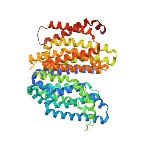
Crystal structure of the MOP flippase MurJ in an inward-facing conformation.

Kuk, A.C.Mashalidis, E.H.Lee, S.Y.

(2017) Nat Struct Mol Biol 24: 171-176

  • DOI: https://doi.org/10.1038/nsmb.3346
  • Primary Citation Related Structures: 
    5T77

  • PubMed Abstract: 

    Peptidoglycan (PG) protects bacteria from osmotic lysis, and its biogenesis is a key antibiotic target. A central step in PG biosynthesis is flipping of the lipid-linked PG precursor lipid II across the cytoplasmic membrane for subsequent incorporation into PG. MurJ, part of the multidrug/oligosaccharidyl-lipid/polysaccharide (MOP) transporter superfamily, was recently shown to carry out this process. However, understanding of how MurJ flips lipid II, and of how MOP transporters operate in general, remains limited due to a lack of structural information. Here we present a crystal structure of MurJ from Thermosipho africanus in an inward-facing conformation at 2.0-Å resolution. A hydrophobic groove is formed by two C-terminal transmembrane helices, which leads into a large central cavity that is mostly cationic. Our studies not only provide the first structural glimpse of MurJ but also suggest that alternating access is important for MurJ function, which may be applicable to other MOP superfamily transporters.


  • Organizational Affiliation
    • Department of Biochemistry, Duke University School of Medicine, Durham, North Carolina, USA.

Macromolecule Content 

  • Total Structure Weight: 69.17 kDa 
  • Atom Count: 4,404 
  • Modeled Residue Count: 467 
  • Deposited Residue Count: 475 
  • Unique protein chains: 1

Macromolecules

Find similar proteins by:|  3D Structure
Entity ID: 1
MoleculeChains  Sequence LengthOrganismDetailsImage
Putative lipid II flippase MurJ475Thermosipho africanus TCF52BMutation(s): 0 
Gene Names: mviNTHA_1814
Membrane Entity: Yes 
UniProt
Find proteins for B7IE18 (Thermosipho africanus (strain TCF52B))
Explore B7IE18 
Go to UniProtKB:  B7IE18
Entity Groups
Sequence Clusters30% Identity50% Identity70% Identity90% Identity95% Identity100% Identity
UniProt GroupB7IE18
Sequence Annotations
Expand
Reference Sequence

Small Molecules

Ligands 7 Unique
IDChains Name / Formula / InChI Key2D Diagram3D Interactions
OLB

Query on OLB


Download:Ideal Coordinates CCD File
P [auth A]
Q [auth A]
R [auth A]
S [auth A]
T [auth A]
P [auth A],
Q [auth A],
R [auth A],
S [auth A],
T [auth A],
U [auth A],
V [auth A]
(2S)-2,3-dihydroxypropyl (9Z)-octadec-9-enoate
C21 H40 O4
RZRNAYUHWVFMIP-QJRAZLAKSA-N
OLC

Query on OLC


Download:Ideal Coordinates CCD File
AA [auth A]
AB [auth A]
BA [auth A]
BB [auth A]
CA [auth A]
AA [auth A],
AB [auth A],
BA [auth A],
BB [auth A],
CA [auth A],
DA [auth A],
EA [auth A],
FA [auth A],
GA [auth A],
HA [auth A],
IA [auth A],
JA [auth A],
KA [auth A],
LA [auth A],
MA [auth A],
NA [auth A],
OA [auth A],
PA [auth A],
QA [auth A],
RA [auth A],
SA [auth A],
TA [auth A],
UA [auth A],
VA [auth A],
W [auth A],
WA [auth A],
X [auth A],
XA [auth A],
Y [auth A],
YA [auth A],
Z [auth A],
ZA [auth A]
(2R)-2,3-dihydroxypropyl (9Z)-octadec-9-enoate
C21 H40 O4
RZRNAYUHWVFMIP-GDCKJWNLSA-N
1PE

Query on 1PE


Download:Ideal Coordinates CCD File
M [auth A],
N [auth A],
O [auth A]
PENTAETHYLENE GLYCOL
C10 H22 O6
JLFNLZLINWHATN-UHFFFAOYSA-N
ZN

Query on ZN


Download:Ideal Coordinates CCD File
G [auth A]ZINC ION
Zn
PTFCDOFLOPIGGS-UHFFFAOYSA-N
CA

Query on CA


Download:Ideal Coordinates CCD File
H [auth A],
I [auth A],
J [auth A]
CALCIUM ION
Ca
BHPQYMZQTOCNFJ-UHFFFAOYSA-N
CL

Query on CL


Download:Ideal Coordinates CCD File
B [auth A],
C [auth A],
D [auth A],
E [auth A],
F [auth A]
CHLORIDE ION
Cl
VEXZGXHMUGYJMC-UHFFFAOYSA-M
NA

Query on NA


Download:Ideal Coordinates CCD File
K [auth A],
L [auth A]
SODIUM ION
Na
FKNQFGJONOIPTF-UHFFFAOYSA-N

Experimental Data & Validation

Experimental Data

  • Method: X-RAY DIFFRACTION
  • Resolution: 2.00 Å
  • R-Value Free:  0.214 (Depositor), 0.216 (DCC) 
  • R-Value Work:  0.189 (Depositor), 0.191 (DCC) 
  • R-Value Observed: 0.190 (Depositor) 
Space Group: C 1 2 1
Unit Cell:
Length ( Å )Angle ( ˚ )
a = 94.163α = 90
b = 99.573β = 112.91
c = 74.66γ = 90
Software Package:
Software NamePurpose
PHENIXrefinement
HKL-2000data reduction
HKL-2000data scaling
PHENIXphasing

Structure Validation

View Full Validation Report


Ligand Structure Quality Assessment 


Entry History 

Deposition Data

Revision History  (Full details and data files)

  • Version 1.0: 2016-12-28
    Type: Initial release
  • Version 1.1: 2017-01-04
    Changes: Database references
  • Version 1.2: 2017-02-15
    Changes: Database references
  • Version 1.3: 2024-03-06
    Changes: Data collection, Database references, Derived calculations