5T67 | pdb_00005t67

x-ray structure of the KijD1 C3-methyltransferase from Actinomadura kijaniata in complex with SAH and dTDP-sugar product


Experimental Data Snapshot

  • Method: X-RAY DIFFRACTION
  • Resolution: 1.60 Å
  • R-Value Free: 
    0.249 (Depositor), 0.257 (DCC) 
  • R-Value Work: 
    0.198 (Depositor), 0.207 (DCC) 
  • R-Value Observed: 
    0.201 (Depositor) 

Starting Model: experimental
View more details

wwPDB Validation 3D Report Full Report

Validation slider image for 5T67

Ligand Structure Quality Assessment 


This is version 1.6 of the entry. See complete history

Literature

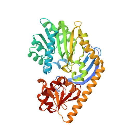
Structural studies on KijD1, a sugar C-3'-methyltransferase.

Dow, G.T.Thoden, J.B.Holden, H.M.

(2016) Protein Sci 25: 2282-2289

  • DOI: https://doi.org/10.1002/pro.3034
  • Primary Citation Related Structures: 
    5T64, 5T67, 5T6B

  • PubMed Abstract: 

    Kijanimicin is an antitumor antibiotic isolated from Actinomadura kijaniata. It is composed of three distinct moieties: a pentacyclic core, a monosaccharide referred to as d-kijanose, and a tetrasaccharide chain composed of l-digitoxose units. d-Kijanose is a highly unusual nitro-containing tetradeoxysugar, which requires at least ten enzymes for its production. Here we describe a structural analysis of one of these enzymes, namely KijD1, which functions as a C-3'-methyltransferase using S-adenosylmethionine as its cofactor. For this investigation, two ternary complexes of KijD1, determined in the presence of S-adenosylhomocysteine (SAH) and dTDP or SAH and dTDP-3-amino-2,3,6-trideoxy-4-keto-3-methyl-d-glucose, were solved to 1.7 or 1.6 Å resolution, respectively. Unexpectedly, these structures, as well as additional biochemical analyses, demonstrated that the quaternary structure of KijD1 is a dimer. Indeed, this is in sharp contrast to that previously observed for the sugar C-3'-methyltransferase isolated from Micromonospora chalcea. By the judicious use of site-directed mutagenesis, it was possible to convert the dimeric form of KijD1 into a monomeric version. The quaternary structure of KijD1 could not have been deduced based solely on bioinformatics approaches, and thus this investigation highlights the continuing need for experimental validation.


  • Organizational Affiliation
    • Department of Biochemistry, University of Wisconsin, Madison, WI, 53706.

Macromolecule Content 

  • Total Structure Weight: 93.98 kDa 
  • Atom Count: 7,249 
  • Modeled Residue Count: 807 
  • Deposited Residue Count: 832 
  • Unique protein chains: 1

Macromolecules

Find similar proteins by:|  3D Structure
Entity ID: 1
MoleculeChains  Sequence LengthOrganismDetailsImage
Sugar 3-C-methyl transferase
A, B
416Actinomadura kijaniataMutation(s): 0 
Gene Names: KijD1
UniProt
Find proteins for B3TMQ9 (Actinomadura kijaniata)
Explore B3TMQ9 
Go to UniProtKB:  B3TMQ9
Entity Groups
Sequence Clusters30% Identity50% Identity70% Identity90% Identity95% Identity100% Identity
UniProt GroupB3TMQ9
Sequence Annotations
Expand
Reference Sequence

Small Molecules

Ligands 6 Unique
IDChains Name / Formula / InChI Key2D Diagram3D Interactions
JHZ

Query on JHZ


Download:Ideal Coordinates CCD File
E [auth A],
N [auth B]
(2R,4S,6R)-4-amino-4,6-dimethyl-5-oxotetrahydro-2H-pyran-2-yl [(2R,3S,5R)-3-hydroxy-5-(5-methyl-2,4-dioxo-3,4-dihydropyrimidin-1(2H)-yl)tetrahydrofuran-2-yl]methyl dihydrogen diphosphate (non-preferred name)
C17 H27 N3 O13 P2
LXNLHPYBLNFMEH-XXXNSVIPSA-N
SAH

Query on SAH


Download:Ideal Coordinates CCD File
D [auth A],
M [auth B]
S-ADENOSYL-L-HOMOCYSTEINE
C14 H20 N6 O5 S
ZJUKTBDSGOFHSH-WFMPWKQPSA-N
ZN

Query on ZN


Download:Ideal Coordinates CCD File
C [auth A],
L [auth B]
ZINC ION
Zn
PTFCDOFLOPIGGS-UHFFFAOYSA-N
EDO

Query on EDO


Download:Ideal Coordinates CCD File
F [auth A],
G [auth A],
H [auth A],
O [auth B]
1,2-ETHANEDIOL
C2 H6 O2
LYCAIKOWRPUZTN-UHFFFAOYSA-N
CL

Query on CL


Download:Ideal Coordinates CCD File
K [auth A]CHLORIDE ION
Cl
VEXZGXHMUGYJMC-UHFFFAOYSA-M
MG

Query on MG


Download:Ideal Coordinates CCD File
I [auth A],
J [auth A]
MAGNESIUM ION
Mg
JLVVSXFLKOJNIY-UHFFFAOYSA-N

Experimental Data & Validation

Experimental Data

  • Method: X-RAY DIFFRACTION
  • Resolution: 1.60 Å
  • R-Value Free:  0.249 (Depositor), 0.257 (DCC) 
  • R-Value Work:  0.198 (Depositor), 0.207 (DCC) 
  • R-Value Observed: 0.201 (Depositor) 
Space Group: P 21 21 21
Unit Cell:
Length ( Å )Angle ( ˚ )
a = 51.088α = 90
b = 108.893β = 90
c = 142.783γ = 90
Software Package:
Software NamePurpose
REFMACrefinement
SAINTdata reduction
SADABSdata scaling
PHASERphasing

Structure Validation

View Full Validation Report


Ligand Structure Quality Assessment 


Entry History 

& Funding Information

Deposition Data


Funding OrganizationLocationGrant Number
National Institutes of Health/National Institute of General Medical Sciences (NIH/NIGMS)United Statesgm115921

Revision History  (Full details and data files)

  • Version 1.0: 2016-09-14
    Type: Initial release
  • Version 1.1: 2016-09-21
    Changes: Database references
  • Version 1.2: 2016-09-28
    Changes: Structure summary
  • Version 1.3: 2016-12-07
    Changes: Database references
  • Version 1.4: 2017-09-27
    Changes: Author supporting evidence, Database references
  • Version 1.5: 2019-12-25
    Changes: Author supporting evidence
  • Version 1.6: 2023-10-04
    Changes: Data collection, Database references, Derived calculations, Refinement description