


Discovery of AMG337: Using structure guided scaffold hydridization to optimize physicochemical properties and target coverage of a MET kinase inhibitor
Boezio, A.A., Peterson, E.A., Harmange, J.-C.To be published.
Experimental Data Snapshot
Starting Model: experimental
View more details
Entity ID: 1 | |||||
|---|---|---|---|---|---|
| Molecule | Chains | Sequence Length | Organism | Details | Image |
| Hepatocyte growth factor receptor | 309 | Homo sapiens | Mutation(s): 0 Gene Names: MET EC: 2.7.10.1 | 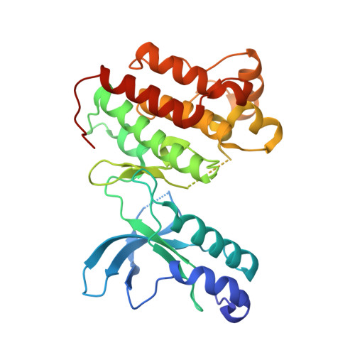 | |
UniProt & NIH Common Fund Data Resources | |||||
PHAROS: P08581 GTEx: ENSG00000105976 | |||||
Entity Groups | |||||
| Sequence Clusters | 30% Identity50% Identity70% Identity90% Identity95% Identity100% Identity | ||||
| UniProt Group | P08581 | ||||
Sequence AnnotationsExpand | |||||
Reference Sequence | |||||
| Ligands 1 Unique | |||||
|---|---|---|---|---|---|
| ID | Chains | Name / Formula / InChI Key | 2D Diagram | 3D Interactions | |
| 75H Download:Ideal Coordinates CCD File | B [auth A] | N-{3-fluoro-4-[(7-methoxyquinolin-4-yl)oxy]phenyl}-1-(2-hydroxy-2-methylpropyl)-5-methyl-3-oxo-2-phenyl-2,3-dihydro-1H-pyrazole-4-carboxamide C31 H29 F N4 O5 UYMSIPINLJNNOU-UHFFFAOYSA-N |  | ||
| Length ( Å ) | Angle ( ˚ ) |
|---|---|
| a = 71.627 | α = 90 |
| b = 82.272 | β = 90 |
| c = 127.139 | γ = 90 |
| Software Name | Purpose |
|---|---|
| SCALEPACK | data scaling |
| REFMAC | refinement |
| PDB_EXTRACT | data extraction |
| DENZO | data reduction |


