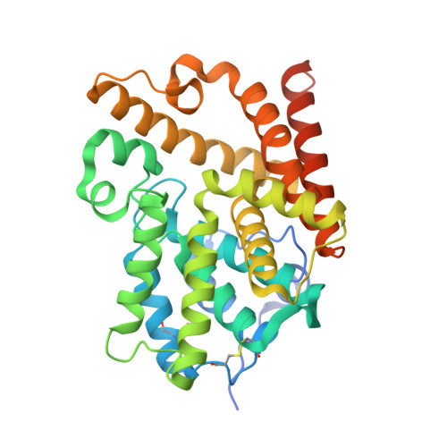
5SFS

CRYSTAL STRUCTURE OF HUMAN PHOSPHODIESTERASE 10 IN COMPLEX WITH n2c(C)c1nc(nn1c(c2)C)CCc3nc(nn3C)N4CC[C@@H](C4)C, micromolar IC50=0.012855


Experimental Data Snapshot

  • Method: X-RAY DIFFRACTION
  • Resolution: 2.24 Å
  • R-Value Free: 0.252 
  • R-Value Work: 0.188 
  • R-Value Observed: 0.191 

Starting Model: other
View more details

wwPDB Validation   3D Report Full Report


Ligand Structure Quality Assessment 

Created with Raphaël 2.3.0Worse 01 BetterLigand structure goodness of fit to experimental dataBest fitted IOUClick on this verticalbar to view details

This is version 1.4 of the entry. See complete history


Literature

A high quality, industrial data set for binding affinity prediction: performance comparison in different early drug discovery scenarios.

Tosstorff, A.Rudolph, M.G.Cole, J.C.Reutlinger, M.Kramer, C.Schaffhauser, H.Nilly, A.Flohr, A.Kuhn, B.

(2022) J Comput Aided Mol Des 36: 753-765

  • DOI: https://doi.org/10.1007/s10822-022-00478-x
  • Primary Citation of Related Structures:  
    5SDU, 5SDV, 5SDW, 5SDX, 5SDY, 5SDZ, 5SE0, 5SE1, 5SE2, 5SE3, 5SE4, 5SE5, 5SE6, 5SE7, 5SE8, 5SE9, 5SEA, 5SEB, 5SEC, 5SED, 5SEE, 5SEF, 5SEG, 5SEH, 5SEI, 5SEJ, 5SEK, 5SEL, 5SEM, 5SEN, 5SEO, 5SEP, 5SEQ, 5SER, 5SES, 5SET, 5SEU, 5SEV, 5SEW, 5SEX, 5SEY, 5SEZ, 5SF0, 5SF1, 5SF2, 5SF3, 5SF4, 5SF5, 5SF6, 5SF7

  • PubMed Abstract: 

    We release a new, high quality data set of 1162 PDE10A inhibitors with experimentally determined binding affinities together with 77 PDE10A X-ray co-crystal structures from a Roche legacy project. This data set is used to compare the performance of different 2D- and 3D-machine learning (ML) as well as empirical scoring functions for predicting binding affinities with high throughput. We simulate use cases that are relevant in the lead optimization phase of early drug discovery. ML methods perform well at interpolation, but poorly in extrapolation scenarios-which are most relevant to a real-world application. Moreover, we find that investing into the docking workflow for binding pose generation using multi-template docking is rewarded with an improved scoring performance. A combination of 2D-ML and 3D scoring using a modified piecewise linear potential shows best overall performance, combining information on the protein environment with learning from existing SAR data.


  • Organizational Affiliation

    Roche Pharma Research and Early Development, Roche Innovation Center Basel, F. Hoffmann-La Roche Ltd., Grenzacherstrasse 124, CH-4070, Basel, Switzerland. a.tosstorff@gmail.com.


Macromolecules
Find similar proteins by:  (by identity cutoff)  |  3D Structure
Entity ID: 1
MoleculeChains Sequence LengthOrganismDetailsImage
cAMP and cAMP-inhibited cGMP 3',5'-cyclic phosphodiesterase 10A
A, B, C, D
343Homo sapiensMutation(s): 0 
Gene Names: PDE10A
EC: 3.1.4.17
UniProt & NIH Common Fund Data Resources
Find proteins for Q9Y233 (Homo sapiens)
Explore Q9Y233 
Go to UniProtKB:  Q9Y233
PHAROS:  Q9Y233
GTEx:  ENSG00000112541 
Entity Groups  
Sequence Clusters30% Identity50% Identity70% Identity90% Identity95% Identity100% Identity
UniProt GroupQ9Y233
Sequence Annotations
Expand
  • Reference Sequence
Small Molecules
Ligands 3 Unique
IDChains Name / Formula / InChI Key2D Diagram3D Interactions
IOU (Subject of Investigation/LOI)
Query on IOU

Download Ideal Coordinates CCD File 
G [auth A],
J [auth B],
M [auth C],
P [auth D]
(4S)-5,8-dimethyl-2-(2-{1-methyl-3-[(3S)-3-methylpyrrolidin-1-yl]-1H-1,2,4-triazol-5-yl}ethyl)[1,2,4]triazolo[1,5-a]pyrazine
C17 H24 N8
ODLMWWMAGVZRPO-UHFFFAOYSA-N
ZN
Query on ZN

Download Ideal Coordinates CCD File 
E [auth A],
H [auth B],
K [auth C],
N [auth D]
ZINC ION
Zn
PTFCDOFLOPIGGS-UHFFFAOYSA-N
MG
Query on MG

Download Ideal Coordinates CCD File 
F [auth A],
I [auth B],
L [auth C],
O [auth D]
MAGNESIUM ION
Mg
JLVVSXFLKOJNIY-UHFFFAOYSA-N
Modified Residues  1 Unique
IDChains TypeFormula2D DiagramParent
CME
Query on CME
A, B, C, D
L-PEPTIDE LINKINGC5 H11 N O3 S2CYS
Experimental Data & Validation

Experimental Data

  • Method: X-RAY DIFFRACTION
  • Resolution: 2.24 Å
  • R-Value Free: 0.252 
  • R-Value Work: 0.188 
  • R-Value Observed: 0.191 
  • Space Group: H 3
Unit Cell:
Length ( Å )Angle ( ˚ )
a = 135.57α = 90
b = 135.57β = 90
c = 235.303γ = 120
Software Package:
Software NamePurpose
XSCALEdata scaling
REFMACrefinement
PDB_EXTRACTdata extraction
XDSdata reduction
XSCALEdata scaling
PHASERphasing

Structure Validation

View Full Validation Report


Ligand Structure Quality Assessment 

Created with Raphaël 2.3.0Worse 01 BetterLigand structure goodness of fit to experimental dataBest fitted IOUClick on this verticalbar to view details

Entry History & Funding Information

Deposition Data


Funding OrganizationLocationGrant Number
F. Hoffmann-La RocheSwitzerland--

Revision History  (Full details and data files)

  • Version 1.0: 2022-10-12
    Type: Initial release
  • Version 1.1: 2022-11-02
    Changes: Database references
  • Version 1.2: 2024-04-03
    Changes: Data collection, Refinement description
  • Version 1.3: 2024-10-09
    Changes: Structure summary
  • Version 1.4: 2024-10-16
    Changes: Structure summary