Binding-Site Purification of Actives (B-SPA) Enables Efficient Large-Scale Progression of Fragment Hits by Combining Multi-Step Array Synthesis With HT Crystallography.
Grosjean, H., Aimon, A., Hassell-Hart, S., Thompson, W., Koekemoer, L., Bennett, J., Bradley, A., Anderson, C., Wild, C., Bradshaw, W.J., FitzGerald, E.A., Krojer, T., Fedorov, O., Biggin, P.C., Spencer, J., von Delft, F.(2025) Angew Chem Int Ed Engl 64: e202424373-e202424373
- PubMed: 39931803 Search on PubMedSearch on PubMed Central
- DOI: https://doi.org/10.1002/anie.202424373
- Primary Citation Related Structures:
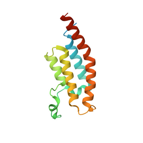
5S8R, 5S8S, 5S8T, 5S8U, 5S8V, 5S8W, 5S8X, 5S8Y, 5S8Z, 5S90, 5S91, 5S92, 5S93, 5S94, 5S95, 5S96, 5S97, 5S98, 5S99, 5S9A, 5S9B, 5S9C, 5S9D, 5S9E, 5S9G, 5S9J, 8BW2, 8BW3, 8BW4 - PubMed Abstract:
Fragment approaches are long-established in target-based ligand discovery, yet their full transformative potential lies dormant because progressing the initial weakly binding hits to potency remains a formidable challenge. The only credible progression paradigm involves multiple cycles of costly conventional design-make-test-analyse medicinal chemistry. We propose an alternative approach to fragment elaboration, namely performing large numbers of parallel and diverse automated multiple step reactions, and evaluating the binding of the crude reaction products by high-throughput protein X-ray crystallography. We show it is effective and low-cost to perform, in parallel, large numbers of non-uniform multi-step reactions, because, even without compound purification, crystallography provides a high-quality readout of binding. This can detect low-level binding of weakly active compounds, which the target binding site extracts directly from crude reaction mixtures. In this proof-of-concept study, we have expanded a fragment hit, from a crystal-based screen of the second bromodomain of pleckstrin homology domain-interacting protein (PHIP(2)), using array synthesis on low-cost robotics. We were able to implement 6 independent multi-step reaction routes of up to 5 steps, attempting the synthesis of 1876 diverse expansions, designs entirely driven by synthetic tractability. The expected product was present in 1108 (59%) crude reaction mixtures, detected by liquid chromatography mass spectrometry (LCMS). 22 individual products were resolved in the crystal structures of crude reaction mixtures added to crystals, providing an initial structure activity relationship map. 19 of these showed binding pose stability, while, through binding instability in the remaining 3 products, we could resolve a stereochemical preference for mixtures containing racemic compounds. One compound showed biochemical potency (IC 50 =34 μM) and affinity (K d =50 μM) after resynthesis. This approach therefore lends itself to routine fragment progression, if coupled with algorithmically guided compound and reaction design and new formalisms for data analysis.
- Diamond Light Source Ltd, Harwell Science and Innovation Campus, OX11 0QX, Didcot, UK.
Organizational Affiliation: