5RHN | pdb_00005rhn

HISTIDINE TRIAD NUCLEOTIDE-BINDING PROTEIN (HINT) FROM RABBIT COMPLEXED WITH 8-BR-AMP


Experimental Data Snapshot

  • Method: X-RAY DIFFRACTION
  • Resolution: 2.31 Å
  • R-Value Free: 
    0.264 (Depositor), 0.243 (DCC) 
  • R-Value Work: 
    0.162 (Depositor), 0.153 (DCC) 
  • R-Value Observed: 
    0.162 (Depositor) 

wwPDB Validation 3D Report Full Report

Validation slider image for 5RHN

Ligand Structure Quality Assessment 


This is version 1.3 of the entry. See complete history

Literature

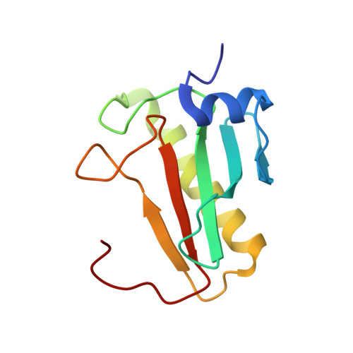
Crystal structures of HINT demonstrate that histidine triad proteins are GalT-related nucleotide-binding proteins.

Brenner, C.Garrison, P.Gilmour, J.Peisach, D.Ringe, D.Petsko, G.A.Lowenstein, J.M.

(1997) Nat Struct Biol 4: 231-238

  • DOI: https://doi.org/10.1038/nsb0397-231
  • Primary Citation Related Structures: 
    3RHN, 4RHN, 5RHN, 6RHN

  • PubMed Abstract: 

    Histidine triad nucleotide-binding protein (HINT), a dimeric purine nucleotide-binding protein from rabbit heart, is a member of the HIT (histidine triad) superfamily which includes HINT homologues and FHIT (HIT protein encoded at the chromosome 3 fragile site) homologues. Crystal structures of HINT-nucleotide complexes demonstrate that the most conserved residues in the superfamily mediate nucleotide binding and that the HIT motif forms part of the phosphate binding loop. Galactose-1-phosphate uridylyltransferase, whose deficiency causes galactosemia, contains tandem HINT domains with the same fold and mode of nucleotide binding as HINT despite having no overall sequence similarity. Features of FHIT, a diadenosine polyphosphate hydrolase and candidate tumour suppressor, are predicted from HINT-nucleotide structures.


  • Organizational Affiliation
    • Department of Biochemistry, Brandeis University, Waltham, Massachusetts 02254, USA. brenner@dada.jci.tju.edu

Macromolecule Content 

  • Total Structure Weight: 13.01 kDa 
  • Atom Count: 953 
  • Modeled Residue Count: 115 
  • Deposited Residue Count: 115 
  • Unique protein chains: 1

Macromolecules

Find similar proteins by:|  3D Structure
Entity ID: 1
MoleculeChains  Sequence LengthOrganismDetailsImage
HISTIDINE TRIAD NUCLEOTIDE-BINDING PROTEIN115Oryctolagus cuniculusMutation(s): 0 
Gene Names: HINT
EC: 3.4.22 (UniProt), 3.9.1 (UniProt)
UniProt
Find proteins for P80912 (Oryctolagus cuniculus)
Explore P80912 
Go to UniProtKB:  P80912
Entity Groups
Sequence Clusters30% Identity50% Identity70% Identity90% Identity95% Identity100% Identity
UniProt GroupP80912
Sequence Annotations
Expand
Reference Sequence

Small Molecules

Ligands 1 Unique
IDChains Name / Formula / InChI Key2D Diagram3D Interactions
8BR

Query on 8BR


Download:Ideal Coordinates CCD File
B [auth A]8-BROMO-ADENOSINE-5'-MONOPHOSPHATE
C10 H13 Br N5 O7 P
DNPIJKNXFSPNNY-UUOKFMHZSA-N

Experimental Data & Validation

Experimental Data

  • Method: X-RAY DIFFRACTION
  • Resolution: 2.31 Å
  • R-Value Free:  0.264 (Depositor), 0.243 (DCC) 
  • R-Value Work:  0.162 (Depositor), 0.153 (DCC) 
  • R-Value Observed: 0.162 (Depositor) 
Space Group: P 43 21 2
Unit Cell:
Length ( Å )Angle ( ˚ )
a = 40.32α = 90
b = 40.32β = 90
c = 143.08γ = 90
Software Package:
Software NamePurpose
X-PLORmodel building
X-PLORrefinement
R-AXISdata reduction
R-AXISdata scaling
X-PLORphasing

Structure Validation

View Full Validation Report


Ligand Structure Quality Assessment 


Entry History 

Deposition Data

Revision History  (Full details and data files)

  • Version 1.0: 1997-06-16
    Type: Initial release
  • Version 1.1: 2008-03-25
    Changes: Version format compliance
  • Version 1.2: 2011-07-13
    Changes: Derived calculations, Version format compliance
  • Version 1.3: 2024-03-06
    Changes: Data collection, Database references, Derived calculations, Other