5OYM | pdb_00005oym

HIV Integrase Binding Domain of Lens Epithelium-Derived Growth Factor


Experimental Data Snapshot

  • Method: X-RAY DIFFRACTION
  • Resolution: 2.05 Å
  • R-Value Free: 
    0.236 (Depositor), 0.241 (DCC) 
  • R-Value Work: 
    0.182 (Depositor), 0.189 (DCC) 
  • R-Value Observed: 
    0.185 (Depositor) 

Starting Model: experimental
View more details

wwPDB Validation 3D Report Full Report

Validation slider image for 5OYM

This is version 1.2 of the entry. See complete history

Literature

Cloning, purification and structure determination of the HIV integrase-binding domain of lens epithelium-derived growth factor.

Hannon, C.Cruz-Migoni, A.Platonova, O.Owen, R.L.Nettleship, J.E.Miller, A.Carr, S.B.Harris, G.Rabbitts, T.H.Phillips, S.E.V.

(2018) Acta Crystallogr F Struct Biol Commun 74: 143-149

  • DOI: https://doi.org/10.1107/S2053230X18001553
  • Primary Citation Related Structures: 
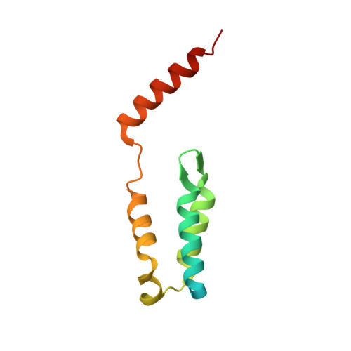
    5OYM

  • PubMed Abstract: 

    Lens epithelium-derived growth factor (LEDGF)/p75 is the dominant binding partner of HIV-1 integrase in human cells. The crystal structure of the HIV integrase-binding domain (IBD) of LEDGF has been determined in the absence of ligand. IBD was overexpressed in Escherichia coli, purified and crystallized by sitting-drop vapour diffusion. X-ray diffraction data were collected at Diamond Light Source to a resolution of 2.05 Å. The crystals belonged to space group P2 1 , with eight polypeptide chains in the asymmetric unit arranged as an unusual octamer composed of four domain-swapped IBD dimers. IBD exists as a mixture of monomers and dimers in concentrated solutions, but the dimers are unlikely to be biologically relevant.


  • Organizational Affiliation
    • Weatherall Institute of Molecular Medicine, University of Oxford, John Radcliffe Hospital, Oxford OX3 9DS, England.

Macromolecule Content 

  • Total Structure Weight: 95.18 kDa 
  • Atom Count: 6,129 
  • Modeled Residue Count: 687 
  • Deposited Residue Count: 888 
  • Unique protein chains: 1

Macromolecules

Find similar proteins by:|  3D Structure
Entity ID: 1
MoleculeChains  Sequence LengthOrganismDetailsImage
PC4 and SFRS1-interacting protein111Homo sapiensMutation(s): 0 
Gene Names: PSIP1DFS70LEDGFPSIP2
UniProt & NIH Common Fund Data Resources
Find proteins for O75475 (Homo sapiens)
Explore O75475 
Go to UniProtKB:  O75475
PHAROS:  O75475
GTEx:  ENSG00000164985 
Entity Groups
Sequence Clusters30% Identity50% Identity70% Identity90% Identity95% Identity100% Identity
UniProt GroupO75475
Sequence Annotations
Expand
Reference Sequence

Experimental Data & Validation

Experimental Data

  • Method: X-RAY DIFFRACTION
  • Resolution: 2.05 Å
  • R-Value Free:  0.236 (Depositor), 0.241 (DCC) 
  • R-Value Work:  0.182 (Depositor), 0.189 (DCC) 
  • R-Value Observed: 0.185 (Depositor) 
Space Group: P 1 21 1
Unit Cell:
Length ( Å )Angle ( ˚ )
a = 71.181α = 90
b = 54.814β = 91.23
c = 117.996γ = 90
Software Package:
Software NamePurpose
REFMACrefinement
XDSdata reduction
SCALAdata scaling
PHASERphasing

Structure Validation

View Full Validation Report


Entry History 

& Funding Information

Deposition Data


Funding OrganizationLocationGrant Number
Medical Research Council (United Kingdom)United KingdomMR/J000612/1
Medical Research Council (United Kingdom)United KingdomMR/K018779/1
BloodwiseUnited Kingdom12051

Revision History  (Full details and data files)

  • Version 1.0: 2018-03-07
    Type: Initial release
  • Version 1.1: 2018-04-18
    Changes: Data collection, Database references
  • Version 1.2: 2024-01-17
    Changes: Data collection, Database references, Refinement description