5OXD | pdb_00005oxd

Complex of a C. perfringens O-GlcNAcase with a fragment hit


Experimental Data Snapshot

  • Method: X-RAY DIFFRACTION
  • Resolution: 2.60 Å
  • R-Value Free: 
    0.221 (Depositor), 0.224 (DCC) 
  • R-Value Work: 
    0.172 (Depositor), 0.180 (DCC) 
  • R-Value Observed: 
    0.174 (Depositor) 

Starting Model: experimental
View more details

wwPDB Validation 3D Report Full Report

Validation slider image for 5OXD

Ligand Structure Quality Assessment 


This is version 1.2 of the entry. See complete history

Literature

O-GlcNAcase Fragment Discovery with Fluorescence Polarimetry.

Borodkin, V.S.Rafie, K.Selvan, N.Aristotelous, T.Navratilova, I.Ferenbach, A.T.van Aalten, D.M.F.

(2018) ACS Chem Biol 13: 1353-1360

  • DOI: https://doi.org/10.1021/acschembio.8b00183
  • Primary Citation Related Structures: 
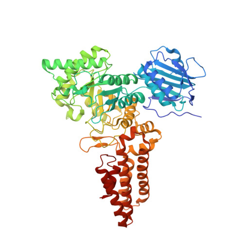
    5OXD

  • PubMed Abstract: 

    The attachment of the sugar N-acetyl-D-glucosamine (GlcNAc) to specific serine and threonine residues on proteins is referred to as protein O-GlcNAcylation. O-GlcNAc transferase (OGT) is the enzyme responsible for carrying out the modification, while O-GlcNAcase (OGA) reverses it. Protein O-GlcNAcylation has been implicated in a wide range of cellular processes including transcription, proteostasis, and stress response. Dysregulation of O-GlcNAc has been linked to diabetes, cancer, and neurodegenerative and cardiovascular disease. OGA has been proposed to be a drug target for the treatment of Alzheimer's and cardiovascular disease given that increased O-GlcNAc levels appear to exert a protective effect. The search for specific, potent, and drug-like OGA inhibitors with bioavailability in the brain is therefore a field of active research, requiring orthogonal high-throughput assay platforms. Here, we describe the synthesis of a novel probe for use in a fluorescence polarization based assay for the discovery of inhibitors of OGA. We show that the probe is suitable for use with both human OGA, as well as the orthologous bacterial counterpart from Clostridium perfringens, CpOGA, and the lysosomal hexosaminidases HexA/B. We structurally characterize CpOGA in complex with a ligand identified from a fragment library screen using this assay. The versatile synthesis procedure could be adapted for making fluorescent probes for the assay of other glycoside hydrolases.

Macromolecule Content 

  • Total Structure Weight: 68.77 kDa 
  • Atom Count: 4,690 
  • Modeled Residue Count: 580 
  • Deposited Residue Count: 590 
  • Unique protein chains: 1

Macromolecules

Find similar proteins by:|  3D Structure
Entity ID: 1
MoleculeChains  Sequence LengthOrganismDetailsImage
O-GlcNAcase NagJ590Clostridium perfringensMutation(s): 0 
Gene Names: nagJCPF_1442
EC: 3.2.1.169 (PDB Primary Data), 3.2.1.52 (PDB Primary Data)
UniProt
Find proteins for Q0TR53 (Clostridium perfringens (strain ATCC 13124 / DSM 756 / JCM 1290 / NCIMB 6125 / NCTC 8237 / Type A))
Explore Q0TR53 
Go to UniProtKB:  Q0TR53
Entity Groups
Sequence Clusters30% Identity50% Identity70% Identity90% Identity95% Identity100% Identity
UniProt GroupQ0TR53
Sequence Annotations
Expand
Reference Sequence

Small Molecules

Ligands 2 Unique
IDChains Name / Formula / InChI Key2D Diagram3D Interactions
B2W

Query on B2W


Download:Ideal Coordinates CCD File
X [auth A]5-(trifluoromethyl)-2,3-dihydro-1~{H}-1,4-diazepine
C6 H7 F3 N2
DQLAMAHCMYJIBW-UHFFFAOYSA-N
CD

Query on CD


Download:Ideal Coordinates CCD File
B [auth A]
C [auth A]
D [auth A]
E [auth A]
F [auth A]
B [auth A],
C [auth A],
D [auth A],
E [auth A],
F [auth A],
G [auth A],
H [auth A],
I [auth A],
J [auth A],
K [auth A],
L [auth A],
M [auth A],
N [auth A],
O [auth A],
P [auth A],
Q [auth A],
R [auth A],
S [auth A],
T [auth A],
U [auth A],
V [auth A],
W [auth A]
CADMIUM ION
Cd
WLZRMCYVCSSEQC-UHFFFAOYSA-N

Experimental Data & Validation

Experimental Data

  • Method: X-RAY DIFFRACTION
  • Resolution: 2.60 Å
  • R-Value Free:  0.221 (Depositor), 0.224 (DCC) 
  • R-Value Work:  0.172 (Depositor), 0.180 (DCC) 
  • R-Value Observed: 0.174 (Depositor) 
Space Group: P 61
Unit Cell:
Length ( Å )Angle ( ˚ )
a = 117.794α = 90
b = 117.794β = 90
c = 147.233γ = 120
Software Package:
Software NamePurpose
REFMACrefinement
XDSdata reduction
Aimlessdata scaling
MOLREPphasing

Structure Validation

View Full Validation Report


Ligand Structure Quality Assessment 


Entry History 

& Funding Information

Deposition Data


Funding OrganizationLocationGrant Number
Wellcome TrustUnited KingdomWT087590MA

Revision History  (Full details and data files)

  • Version 1.0: 2018-05-16
    Type: Initial release
  • Version 1.1: 2018-05-30
    Changes: Data collection, Database references
  • Version 1.2: 2024-01-17
    Changes: Data collection, Database references, Derived calculations, Refinement description