5OST | pdb_00005ost

Beta-glucosidase from Thermoanaerobacterium xylolyticum GH116 in complex with Gluco-1H-imidazole


Experimental Data Snapshot

  • Method: X-RAY DIFFRACTION
  • Resolution: 2.10 Å
  • R-Value Free: 
    0.251 (Depositor), 0.260 (DCC) 
  • R-Value Work: 
    0.200 (Depositor), 0.209 (DCC) 
  • R-Value Observed: 
    0.202 (Depositor) 

Starting Model: experimental
View more details

wwPDB Validation 3D Report Full Report

Validation slider image for 5OST

Ligand Structure Quality Assessment 


This is version 1.3 of the entry. See complete history

Literature

Gluco-1 H-imidazole: A New Class of Azole-Type beta-Glucosidase Inhibitor.

Schroder, S.P.Wu, L.Artola, M.Hansen, T.Offen, W.A.Ferraz, M.J.Li, K.Y.Aerts, J.M.F.G.van der Marel, G.A.Codee, J.D.C.Davies, G.J.Overkleeft, H.S.

(2018) J Am Chem Soc 140: 5045-5048

  • DOI: https://doi.org/10.1021/jacs.8b02399
  • Primary Citation Related Structures: 
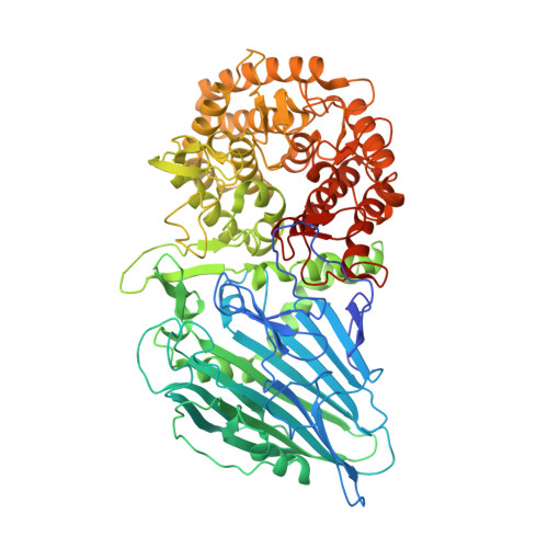
    5OSS, 5OST

  • PubMed Abstract: 

    Gluco-azoles competitively inhibit glucosidases by transition-state mimicry and their ability to interact with catalytic acid residues in glucosidase active sites. We noted that no azole-type inhibitors described, to date, possess a protic nitrogen characteristic for 1 H-imidazoles. Here, we present gluco-1 H-imidazole, a gluco-azole bearing a 1 H-imidazole fused to a glucopyranose-configured cyclitol core, and three close analogues as new glucosidase inhibitors. All compounds inhibit human retaining β-glucosidase, GBA1, with the most potent ones inhibiting this enzyme (deficient in Gaucher disease) on a par with glucoimidazole. None inhibit glucosylceramide synthase, cytosolic β-glucosidase GBA2 or α-glucosidase GAA. Structural, physical and computational studies provide first insights into the binding mode of this conceptually new class of retaining β-glucosidase inhibitors.


  • Organizational Affiliation
    • Department of Chemistry, York Structural Biology Laboratory , University of York , Heslington, York YO10 5DD , United Kingdom.

Macromolecule Content 

  • Total Structure Weight: 92.71 kDa 
  • Atom Count: 6,431 
  • Modeled Residue Count: 762 
  • Deposited Residue Count: 799 
  • Unique protein chains: 1

Macromolecules

Find similar proteins by:|  3D Structure
Entity ID: 1
MoleculeChains  Sequence LengthOrganismDetailsImage
Glucosylceramidase799Thermoanaerobacterium xylanolyticum LX-11Mutation(s): 0 
Gene Names: Thexy_2211
EC: 3.2.1.45
UniProt
Find proteins for F6BL85 (Thermoanaerobacterium xylanolyticum (strain ATCC 49914 / DSM 7097 / LX-11))
Explore F6BL85 
Go to UniProtKB:  F6BL85
Entity Groups
Sequence Clusters30% Identity50% Identity70% Identity90% Identity95% Identity100% Identity
UniProt GroupF6BL85
Sequence Annotations
Expand
Reference Sequence

Small Molecules

Ligands 4 Unique
IDChains Name / Formula / InChI Key2D Diagram3D Interactions
AEZ

Query on AEZ


Download:Ideal Coordinates CCD File
P [auth A](4~{S},5~{S},6~{R},7~{R})-7-(hydroxymethyl)-4,5,6,7-tetrahydro-1~{H}-benzimidazole-4,5,6-triol
C8 H12 N2 O4
MTUBISADCTVQLI-FBXHKNTESA-N
EDO

Query on EDO


Download:Ideal Coordinates CCD File
C [auth A]
D [auth A]
E [auth A]
F [auth A]
G [auth A]
C [auth A],
D [auth A],
E [auth A],
F [auth A],
G [auth A],
H [auth A],
I [auth A],
J [auth A]
1,2-ETHANEDIOL
C2 H6 O2
LYCAIKOWRPUZTN-UHFFFAOYSA-N
CA

Query on CA


Download:Ideal Coordinates CCD File
B [auth A]CALCIUM ION
Ca
BHPQYMZQTOCNFJ-UHFFFAOYSA-N
CL

Query on CL


Download:Ideal Coordinates CCD File
K [auth A],
L [auth A],
M [auth A],
N [auth A],
O [auth A]
CHLORIDE ION
Cl
VEXZGXHMUGYJMC-UHFFFAOYSA-M

Experimental Data & Validation

Experimental Data

  • Method: X-RAY DIFFRACTION
  • Resolution: 2.10 Å
  • R-Value Free:  0.251 (Depositor), 0.260 (DCC) 
  • R-Value Work:  0.200 (Depositor), 0.209 (DCC) 
  • R-Value Observed: 0.202 (Depositor) 
Space Group: P 21 21 2
Unit Cell:
Length ( Å )Angle ( ˚ )
a = 177.617α = 90
b = 54.09β = 90
c = 83.559γ = 90
Software Package:
Software NamePurpose
REFMACrefinement
DIALSdata reduction
Aimlessdata scaling
REFMACphasing

Structure Validation

View Full Validation Report


Ligand Structure Quality Assessment 


Entry History 

& Funding Information

Deposition Data


Funding OrganizationLocationGrant Number
European Research CouncilUnited KingdomERC-2012-AdG-322942

Revision History  (Full details and data files)

  • Version 1.0: 2018-04-11
    Type: Initial release
  • Version 1.1: 2018-04-25
    Changes: Data collection, Database references, Derived calculations, Structure summary
  • Version 1.2: 2018-05-09
    Changes: Data collection, Structure summary
  • Version 1.3: 2024-01-17
    Changes: Data collection, Database references, Refinement description