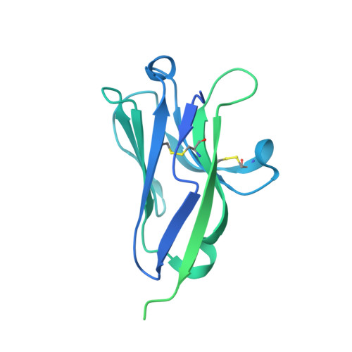
Atomic Structure of the Murine Norovirus Protruding Domain and Soluble CD300lf Receptor Complex.
Kilic, T., Koromyslova, A., Malak, V., Hansman, G.S.(2018) J Virol 92
- PubMed: 29563286 Search on PubMedSearch on PubMed Central
- DOI: https://doi.org/10.1128/JVI.00413-18
- Primary Citation Related Structures:
5OR7 - PubMed Abstract:
Human noroviruses are the leading cause of acute gastroenteritis in humans. Noroviruses also infect animals, such as cows, mice, cats, and dogs. How noroviruses bind and enter host cells is still incompletely understood. Recently, the type I transmembrane protein CD300lf was identified as the murine norovirus receptor, yet it is unclear how the virus capsid and receptor interact at the molecular level. In this study, we determined the X-ray crystal structure of the soluble CD300lf (sCD300lf) and the murine norovirus capsid protruding domain complex at a 2.05-Å resolution. We found that the sCD300lf-binding site is located on the topside of the protruding domain and involves a network of hydrophilic and hydrophobic interactions. sCD300lf locked nicely into a complementary cavity on the protruding domain that is additionally coordinated with a positive surface charge on sCD300lf and a negative surface charge on the protruding domain. Five of six protruding domain residues interacting with sCD300lf were maintained between different murine norovirus strains, suggesting that sCD300lf was capable of binding to a highly conserved pocket. Moreover, a sequence alignment with other CD300 paralogs showed that the sCD300lf-interacting residues were partially conserved in CD300ld but variable in other CD300 family members, consistent with previously reported infection selectivity. Overall, these data provide insights into how a norovirus engages a protein receptor and will be important for a better understanding of selective recognition and norovirus attachment and entry mechanisms. IMPORTANCE Noroviruses exhibit exquisite host range specificity due to species-specific interactions between the norovirus capsid protein and host molecules. Given this strict host range restriction, it has been unclear how the viruses are maintained within a species between relatively sporadic epidemics. While much data demonstrate that noroviruses can interact with carbohydrates, recent work has shown that expression of the protein CD300lf is both necessary and sufficient for murine norovirus infection of mice and binding of the virus to permissive cells. Importantly, the expression of this murine protein by human cells renders them fully permissive for murine norovirus infection, indicating that at least in this case, host range restriction is determined by molecular events that control receptor binding and entry. Defining the atomic-resolution interactions between the norovirus capsid protein and its cognate receptor is essential for a molecular understanding of host-range restriction and norovirus tropism.
- Schaller Research Group at the University of Heidelberg and the DKFZ, Heidelberg, Germany.
Organizational Affiliation: