5OP9 | pdb_00005op9

The crystal structure of P450 CYP121 in complex with lead compound 7e


Experimental Data Snapshot

  • Method: X-RAY DIFFRACTION
  • Resolution: 1.46 Å
  • R-Value Free: 
    0.173 (Depositor), 0.183 (DCC) 
  • R-Value Work: 
    0.147 (Depositor), 0.159 (DCC) 
  • R-Value Observed: 
    0.148 (Depositor) 

Starting Model: other
View more details

wwPDB Validation 3D Report Full Report

Validation slider image for 5OP9

Ligand Structure Quality Assessment 


This is version 1.1 of the entry. See complete history

Literature

Novel Aryl Substituted Pyrazoles as Small Molecule Inhibitors of Cytochrome P450 CYP121A1: Synthesis and Antimycobacterial Evaluation.

Taban, I.M.Elshihawy, H.E.A.E.Torun, B.Zucchini, B.Williamson, C.J.Altuwairigi, D.Ngu, A.S.T.McLean, K.J.Levy, C.W.Sood, S.Marino, L.B.Munro, A.W.de Carvalho, L.P.S.Simons, C.

(2017) J Med Chem 60: 10257-10267

  • DOI: https://doi.org/10.1021/acs.jmedchem.7b01562
  • Primary Citation Related Structures: 
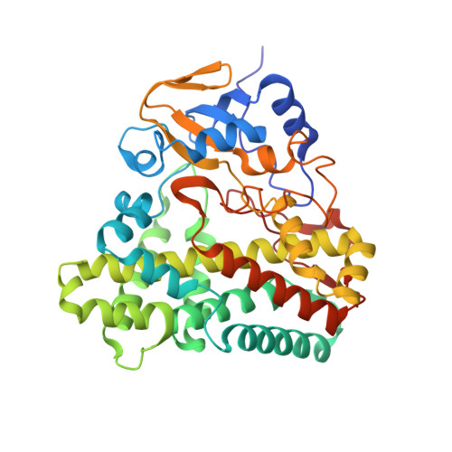
    5O4K, 5O4L, 5OP9, 5OPA

  • PubMed Abstract: 

    Three series of biarylpyrazole imidazole and triazoles are described, which vary in the linker between the biaryl pyrazole and imidazole/triazole group. The imidazole and triazole series with the short -CH 2 - linker displayed promising antimycobacterial activity, with the imidazole-CH 2 - series (7) showing low MIC values (6.25-25 μg/mL), which was also influenced by lipophilicity. Extending the linker to -C(O)NH(CH 2 ) 2 - resulted in a loss of antimycobacterial activity. The binding affinity of the compounds with CYP121A1 was determined by UV-visible optical titrations with K D values of 2.63, 35.6, and 290 μM, respectively, for the tightest binding compounds 7e, 8b, and 13d from their respective series. Both binding affinity assays and docking studies of the CYP121A1 inhibitors suggest type II indirect binding through interstitial water molecules, with key binding residues Thr77, Val78, Val82, Val83, Met86, Ser237, Gln385, and Arg386, comparable with the binding interactions observed with fluconazole and the natural substrate dicyclotyrosine.


  • Organizational Affiliation
    • School of Pharmacy & Pharmaceutical Sciences, Cardiff University , King Edward VII Avenue, Cardiff CF10 3NB, U.K.

Macromolecule Content 

  • Total Structure Weight: 44.83 kDa 
  • Atom Count: 3,833 
  • Modeled Residue Count: 394 
  • Deposited Residue Count: 396 
  • Unique protein chains: 1

Macromolecules

Find similar proteins by:|  3D Structure
Entity ID: 1
MoleculeChains  Sequence LengthOrganismDetailsImage
Mycocyclosin synthase396Mycobacterium tuberculosisMutation(s): 0 
Gene Names: cyp121MT2336
EC: 1.14.21.9 (PDB Primary Data), 1.14.19.70 (UniProt)
UniProt
Find proteins for P9WPP7 (Mycobacterium tuberculosis (strain ATCC 25618 / H37Rv))
Explore P9WPP7 
Go to UniProtKB:  P9WPP7
Entity Groups
Sequence Clusters30% Identity50% Identity70% Identity90% Identity95% Identity100% Identity
UniProt GroupP9WPP7
Sequence Annotations
Expand
Reference Sequence

Small Molecules

Ligands 3 Unique
IDChains Name / Formula / InChI Key2D Diagram3D Interactions
HEM

Query on HEM


Download:Ideal Coordinates CCD File
E [auth A]PROTOPORPHYRIN IX CONTAINING FE
C34 H32 Fe N4 O4
KABFMIBPWCXCRK-RGGAHWMASA-L
BZ6
(Subject of Investigation/LOI)

Query on BZ6


Download:Ideal Coordinates CCD File
F [auth A]4-(imidazol-1-ylmethyl)-3-(4-methoxyphenyl)-1-phenyl-pyrazole
C20 H18 N4 O
CZOCKHTYPCJURT-UHFFFAOYSA-N
SO4

Query on SO4


Download:Ideal Coordinates CCD File
B [auth A]
C [auth A]
D [auth A]
G [auth A]
H [auth A]
B [auth A],
C [auth A],
D [auth A],
G [auth A],
H [auth A],
I [auth A]
SULFATE ION
O4 S
QAOWNCQODCNURD-UHFFFAOYSA-L

Experimental Data & Validation

Experimental Data

  • Method: X-RAY DIFFRACTION
  • Resolution: 1.46 Å
  • R-Value Free:  0.173 (Depositor), 0.183 (DCC) 
  • R-Value Work:  0.147 (Depositor), 0.159 (DCC) 
  • R-Value Observed: 0.148 (Depositor) 
Space Group: P 65 2 2
Unit Cell:
Length ( Å )Angle ( ˚ )
a = 77.507α = 90
b = 77.507β = 90
c = 264.353γ = 120
Software Package:
Software NamePurpose
PHENIXrefinement
XDSdata reduction
XDSdata scaling
PHASERphasing

Structure Validation

View Full Validation Report


Ligand Structure Quality Assessment 


Entry History 

Deposition Data

  • Released Date: 2018-03-28 
  • Deposition Author(s): Levy, C.W.

Revision History  (Full details and data files)

  • Version 1.0: 2018-03-28
    Type: Initial release
  • Version 1.1: 2024-05-01
    Changes: Data collection, Database references, Refinement description