A folding nucleus and minimal ATP binding domain of Hsp70 identified by single-molecule force spectroscopy.
Bauer, D., Meinhold, S., Jakob, R.P., Stigler, J., Merkel, U., Maier, T., Rief, M., Zoldak, G.(2018) Proc Natl Acad Sci U S A 115: 4666-4671
- PubMed: 29669923 Search on PubMedSearch on PubMed Central
- DOI: https://doi.org/10.1073/pnas.1716899115
- Primary Citation Related Structures:
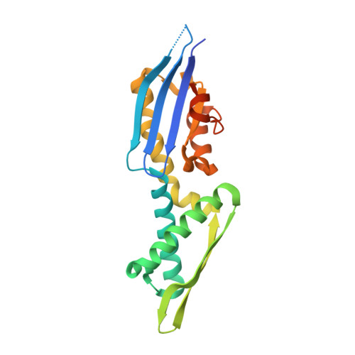
5OOW - PubMed Abstract:
The folding pathways of large proteins are complex, with many of them requiring the aid of chaperones and others folding spontaneously. Along the folding pathways, partially folded intermediates are frequently populated; their role in the driving of the folding process is unclear. The structures of these intermediates are generally not amenable to high-resolution structural techniques because of their transient nature. Here we employed single-molecule force measurements to scrutinize the hierarchy of intermediate structures along the folding pathway of the nucleotide binding domain (NBD) of Escherichia coli Hsp70 DnaK. DnaK-NBD is a member of the sugar kinase superfamily that includes Hsp70s and the cytoskeletal protein actin. Using optical tweezers, a stable nucleotide-binding competent en route folding intermediate comprising lobe II residues (183-383) was identified as a critical checkpoint for productive folding. We obtained a structural snapshot of this folding intermediate that shows native-like conformation. To assess the fundamental role of folded lobe II for efficient folding, we turned our attention to yeast mitochondrial NBD, which does not fold without a dedicated chaperone. After replacing the yeast lobe II residues with stable E. coli lobe II, the obtained chimeric protein showed native-like ATPase activity and robust folding into the native state, even in the absence of chaperone. In summary, lobe II is a stable nucleotide-binding competent folding nucleus that is the key to time-efficient folding and possibly resembles a common ancestor domain. Our findings provide a conceptual framework for the folding pathways of other members of this protein superfamily.
- Physik Department E22, Technische Universität München, 85748 Garching, Germany.
Organizational Affiliation: