5OL4 | pdb_00005ol4

1.28 A resolution of Sporosarcina pasteurii urease inhibited in the presence of NBPT


Experimental Data Snapshot

  • Method: X-RAY DIFFRACTION
  • Resolution: 1.28 Å
  • R-Value Free: 
    0.137 (Depositor), 0.136 (DCC) 
  • R-Value Work: 
    0.119 (Depositor), 0.119 (DCC) 
  • R-Value Observed: 
    0.120 (Depositor) 

Starting Model: experimental
View more details

wwPDB Validation 3D Report Full Report

Validation slider image for 5OL4

This is version 1.1 of the entry. See complete history

Literature

Urease Inhibition in the Presence of N-(n-Butyl)thiophosphoric Triamide, a Suicide Substrate: Structure and Kinetics.

Mazzei, L.Cianci, M.Contaldo, U.Musiani, F.Ciurli, S.

(2017) Biochemistry 56: 5391-5404

  • DOI: https://doi.org/10.1021/acs.biochem.7b00750
  • Primary Citation Related Structures: 
    5OL4

  • PubMed Abstract: 

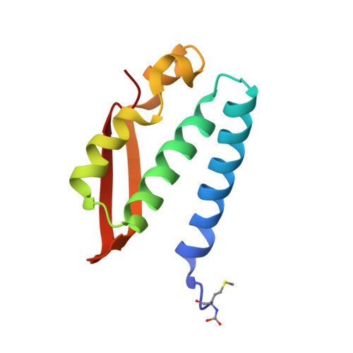
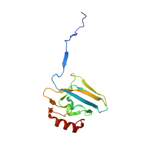
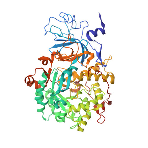
    The nickel-dependent enzyme urease is a virulence factor for a large number of pathogenic and antibiotic-resistant bacteria, as well as a negative factor for the efficiency of soil nitrogen fertilization for crop production. The use of urease inhibitors to offset these effects requires knowledge, at a molecular level, of their mode of action. The 1.28 Å resolution structure of the enzyme-inhibitor complex obtained upon incubation of Sporosarcina pasteurii urease with N-(n-butyl)thiophosphoric triamide (NBPT), a molecule largely utilized in agriculture, reveals the presence of the monoamidothiophosphoric acid (MATP) moiety, obtained upon enzymatic hydrolysis of the diamide derivative of NBPT (NBPD) to yield n-butyl amine. MATP is bound to the two Ni(II) ions in the active site of urease using a μ 2 -bridging O atom and terminally bound O and NH 2 groups, with the S atom of the thiophosphoric amide pointing away from the metal center. The mobile flap modulating the size of the active site cavity is found in the closed conformation. Docking calculations suggest that the interaction between urease in the open flap conformation and NBPD involves a role for the conserved αArg339 in capturing and orienting the inhibitor prior to flap closure. Calorimetric and spectrophotometric determinations of the kinetic parameters of this inhibition indicate the occurrence of a reversible slow inhibition mode of action, characterized, for both bacterial and plant ureases, by a very small value of the dissociation constant of the urease-MATP complex. No need to convert NBPT to its oxo derivative NBPTO, as previously proposed, is necessary for urease inhibition.


  • Organizational Affiliation
    • Laboratory of Bioinorganic Chemistry, Department of Pharmacy and Biotechnology, University of Bologna , Bologna, Italy.

Macromolecule Content 

  • Total Structure Weight: 87.81 kDa 
  • Atom Count: 7,202 
  • Modeled Residue Count: 792 
  • Deposited Residue Count: 792 
  • Unique protein chains: 3

Macromolecules

Find similar proteins by:|  3D Structure
Entity ID: 1
MoleculeChains  Sequence LengthOrganismDetailsImage
Urease subunit gamma100Sporosarcina pasteuriiMutation(s): 0 
EC: 3.5.1.5
UniProt
Find proteins for P41022 (Sporosarcina pasteurii)
Explore P41022 
Go to UniProtKB:  P41022
Entity Groups
Sequence Clusters30% Identity50% Identity70% Identity90% Identity95% Identity100% Identity
UniProt GroupP41022
Sequence Annotations
Expand
Reference Sequence
Find similar proteins by:|  3D Structure
Entity ID: 2
MoleculeChains  Sequence LengthOrganismDetailsImage
Urease subunit beta122Sporosarcina pasteuriiMutation(s): 0 
EC: 3.5.1.5
UniProt
Find proteins for P41021 (Sporosarcina pasteurii)
Explore P41021 
Go to UniProtKB:  P41021
Entity Groups
Sequence Clusters30% Identity50% Identity70% Identity90% Identity95% Identity100% Identity
UniProt GroupP41021
Sequence Annotations
Expand
Reference Sequence
Find similar proteins by:|  3D Structure
Entity ID: 3
MoleculeChains  Sequence LengthOrganismDetailsImage
Urease subunit alpha570Sporosarcina pasteuriiMutation(s): 0 
EC: 3.5.1.5
UniProt
Find proteins for P41020 (Sporosarcina pasteurii)
Explore P41020 
Go to UniProtKB:  P41020
Entity Groups
Sequence Clusters30% Identity50% Identity70% Identity90% Identity95% Identity100% Identity
UniProt GroupP41020
Sequence Annotations
Expand
Reference Sequence

Small Molecules

Ligands 4 Unique
IDChains Name / Formula / InChI Key2D Diagram3D Interactions
9XN
(Subject of Investigation/LOI)

Query on 9XN


Download:Ideal Coordinates CCD File
Z [auth C]Phosphoramidothioic O,O-acid
H4 N O2 P S
RJBIAAZJODIFHR-UHFFFAOYSA-N
SO4

Query on SO4


Download:Ideal Coordinates CCD File
N [auth B],
X [auth C],
Y [auth C]
SULFATE ION
O4 S
QAOWNCQODCNURD-UHFFFAOYSA-L
EDO

Query on EDO


Download:Ideal Coordinates CCD File
D [auth A]
E [auth A]
F [auth A]
G [auth A]
H [auth A]
D [auth A],
E [auth A],
F [auth A],
G [auth A],
H [auth A],
I [auth A],
J [auth A],
K [auth B],
L [auth B],
M [auth B],
Q [auth C],
R [auth C],
S [auth C],
T [auth C],
U [auth C],
V [auth C],
W [auth C]
1,2-ETHANEDIOL
C2 H6 O2
LYCAIKOWRPUZTN-UHFFFAOYSA-N
NI

Query on NI


Download:Ideal Coordinates CCD File
O [auth C],
P [auth C]
NICKEL (II) ION
Ni
VEQPNABPJHWNSG-UHFFFAOYSA-N
Modified Residues  2 Unique
IDChains TypeFormula2D DiagramParent
CXM
Query on CXM
A
L-PEPTIDE LINKINGC6 H11 N O4 SMET
KCX
Query on KCX
C
L-PEPTIDE LINKINGC7 H14 N2 O4LYS

Experimental Data & Validation

Experimental Data

  • Method: X-RAY DIFFRACTION
  • Resolution: 1.28 Å
  • R-Value Free:  0.137 (Depositor), 0.136 (DCC) 
  • R-Value Work:  0.119 (Depositor), 0.119 (DCC) 
  • R-Value Observed: 0.120 (Depositor) 
Space Group: P 63 2 2
Unit Cell:
Length ( Å )Angle ( ˚ )
a = 131.742α = 90
b = 131.742β = 90
c = 188.93γ = 120
Software Package:
Software NamePurpose
REFMACrefinement
XDSdata reduction
Aimlessdata scaling
MOLREPphasing

Structure Validation

View Full Validation Report


Entry History 

Deposition Data

Revision History  (Full details and data files)

  • Version 1.0: 2018-03-07
    Type: Initial release
  • Version 1.1: 2024-01-17
    Changes: Data collection, Database references, Derived calculations, Refinement description