5OEW | pdb_00005oew

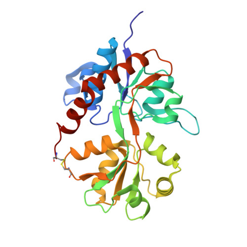
Crystal structure of the GluA2 ligand-binding domain (S1S2J) in complex with glutamate and positive allosteric modulator BPAM538


Experimental Data Snapshot

  • Method: X-RAY DIFFRACTION
  • Resolution: 2.00 Å
  • R-Value Free: 
    0.189 (Depositor), 0.196 (DCC) 
  • R-Value Work: 
    0.167 (Depositor), 0.176 (DCC) 
  • R-Value Observed: 
    0.168 (Depositor) 

Starting Model: experimental
View more details

wwPDB Validation 3D Report Full Report

Validation slider image for 5OEW

Ligand Structure Quality Assessment 


This is version 1.3 of the entry. See complete history. 

Literature

7-Phenoxy-Substituted 3,4-Dihydro-2H-1,2,4-benzothiadiazine 1,1-Dioxides as Positive Allosteric Modulators of alpha-Amino-3-hydroxy-5-methyl-4-isoxazolepropionic Acid (AMPA) Receptors with Nanomolar Potency.

Goffin, E., Drapier, T., Larsen, A.P., Geubelle, P., Ptak, C.P., Laulumaa, S., Rovinskaja, K., Gilissen, J., Tullio, P., Olsen, L., Frydenvang, K., Pirotte, B., Hanson, J., Oswald, R.E., Kastrup, J.S., Francotte, P.

(2018) J Med Chem 61: 251-264

  • DOI: https://doi.org/10.1021/acs.jmedchem.7b01323
  • Primary Citation Related Structures: 
    5O9A, 5OEW

  • PubMed Abstract: 

    We report here the synthesis of 7-phenoxy-substituted 3,4-dihydro-2H-1,2,4-benzothiadiazine 1,1-dioxides and their evaluation as AMPA receptor positive allosteric modulators (AMPApams). The impact of substitution on the phenoxy ring and on the nitrogen atom at the 4-position was examined. At GluA2(Q) expressed in HEK293 cells (calcium flux experiment), the most potent compound was 11m (4-cyclopropyl-7-(3-methoxyphenoxy)-3,4-dihydro-2H-1,2,4-benzothiadiazine 1,1-dioxide, EC 50 = 2.0 nM). The Hill coefficient in the screening and the shape of the dimerization curve in small-angle X-ray scattering (SAXS) experiments using isolated GluA2 ligand-binding domain (GluA2-LBD) are consistent with binding of one molecule of 11m per dimer interface, contrary to most benzothiadiazine dioxides developed to date. This observation was confirmed by the X-ray structure of 11m bound to GluA2-LBD and by NMR. This is the first benzothiadiazine dioxide AMPApam to reach the nanomolar range.


  • Organizational Affiliation: 
    • Laboratory of Medicinal Chemistry, Center for Interdisciplinary Research on Medicines (CIRM), University of Liège , Quartier Hôpital B36 Av. Hippocrate 15 B-4000 Liège, Belgium.

Macromolecule Content 

  • Total Structure Weight: 90.32 kDa 
  • Atom Count: 6,806 
  • Modeled Residue Count: 781 
  • Deposited Residue Count: 792 
  • Unique protein chains: 1

Macromolecules

Find similar proteins by:|  3D Structure
Entity ID: 1
MoleculeChains  Sequence LengthOrganismDetailsImage
Glutamate receptor 2
A, B, C
264Rattus norvegicusMutation(s): 0 
Gene Names: Gria2, Glur2
UniProt
Find proteins for P19491 (Rattus norvegicus)
Explore P19491 
Go to UniProtKB:  P19491
Entity Groups
Sequence Clusters30% Identity50% Identity70% Identity90% Identity95% Identity100% Identity
UniProt GroupP19491
Sequence Annotations
Expand
Reference Sequence

Small Molecules

Ligands 4 Unique
IDChains Name / Formula / InChI Key2D Diagram3D Interactions
9TE
(Subject of Investigation/LOI)

Query on 9TE


Download:Ideal Coordinates CCD File
I [auth B],
R [auth C]
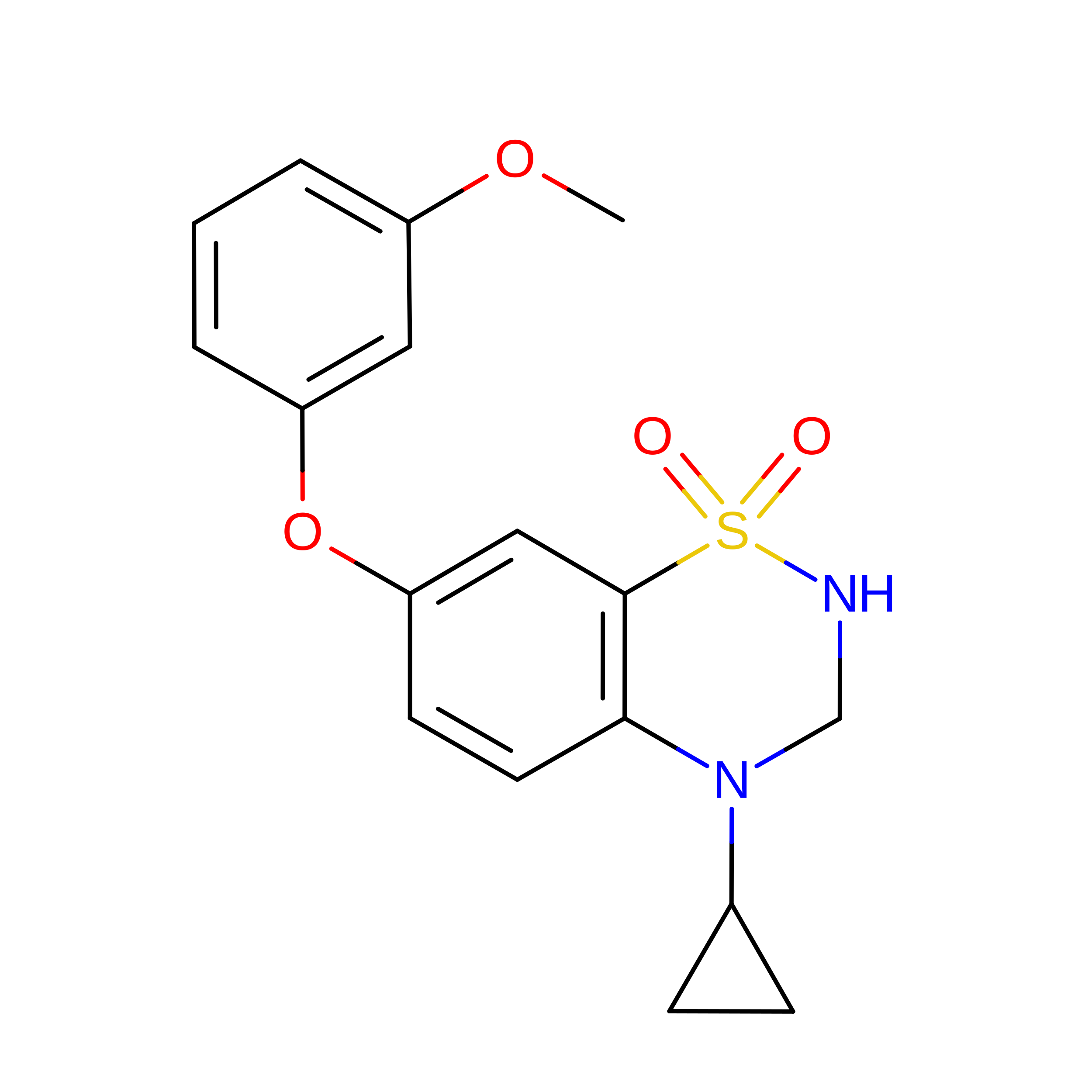
4-cyclopropyl-7-(3-methoxyphenoxy)-2,3-dihydro-1$l^{6},2,4-benzothiadiazine 1,1-dioxide
C17 H18 N2 O4 S
CTMHATKTIDUKBM-UHFFFAOYSA-N
GLU

Query on GLU


Download:Ideal Coordinates CCD File
D [auth A],
J [auth B],
S [auth C]
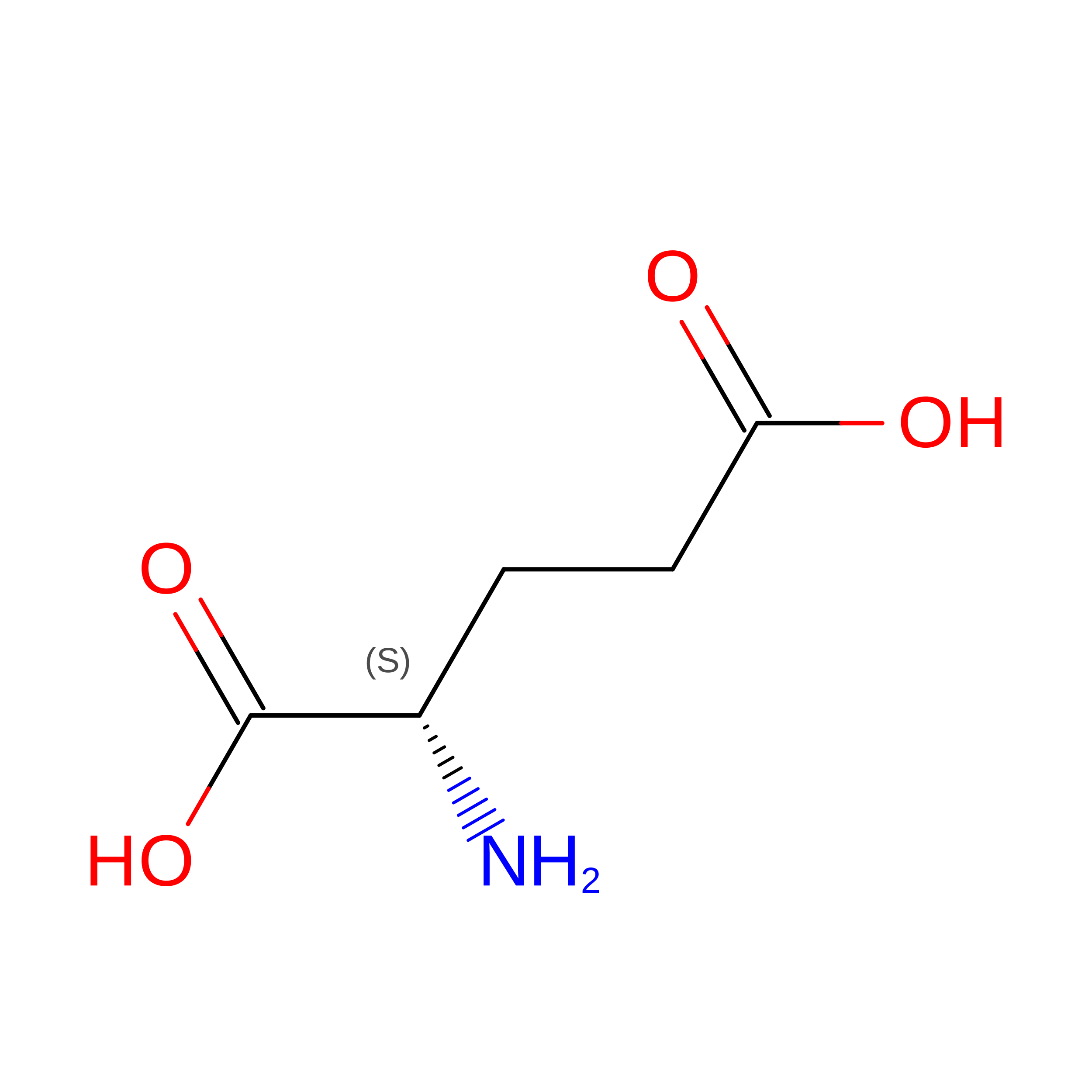
GLUTAMIC ACID
C5 H9 N O4
WHUUTDBJXJRKMK-VKHMYHEASA-N
ZN

Query on ZN


Download:Ideal Coordinates CCD File
AA [auth C]
E [auth A]
F [auth A]
G [auth A]
K [auth B]
AA [auth C],
E [auth A],
F [auth A],
G [auth A],
K [auth B],
L [auth B],
M [auth B],
N [auth B],
O [auth B],
P [auth B],
T [auth C],
U [auth C],
V [auth C],
W [auth C],
X [auth C],
Y [auth C],
Z [auth C]
ZINC ION
Zn
PTFCDOFLOPIGGS-UHFFFAOYSA-N
ACT

Query on ACT


Download:Ideal Coordinates CCD File
BA [auth C],
CA [auth C],
H [auth A],
Q [auth B]
ACETATE ION
C2 H3 O2
QTBSBXVTEAMEQO-UHFFFAOYSA-M

Experimental Data & Validation

Experimental Data

  • Method: X-RAY DIFFRACTION
  • Resolution: 2.00 Å
  • R-Value Free:  0.189 (Depositor), 0.196 (DCC) 
  • R-Value Work:  0.167 (Depositor), 0.176 (DCC) 
  • R-Value Observed: 0.168 (Depositor) 
Space Group: P 21 21 2
Unit Cell:
Length ( Å )Angle ( ˚ )
a = 114.702α = 90
b = 164.374β = 90
c = 47.328γ = 90
Software Package:
Software NamePurpose
SCALAdata scaling
PHASERphasing
PHENIXrefinement
PDB_EXTRACTdata extraction
XDSdata reduction

Structure Validation

View Full Validation Report


Ligand Structure Quality Assessment 


Entry History 

Deposition Data

Revision History  (Full details and data files)

  • Version 1.0: 2018-01-03
    Type: Initial release
  • Version 1.1: 2018-01-24
    Changes: Database references
  • Version 1.2: 2024-01-17
    Changes: Data collection, Database references, Derived calculations, Refinement description
  • Version 1.3: 2024-11-13
    Changes: Structure summary