5OBT | pdb_00005obt

Fully activated A. thaliana legumain isoform gamma in complex with Ac-YVAD-CMK


Experimental Data Snapshot

  • Method: X-RAY DIFFRACTION
  • Resolution: 1.50 Å
  • R-Value Free: 
    0.182 (Depositor), 0.182 (DCC) 
  • R-Value Work: 
    0.152 (Depositor), 0.153 (DCC) 
  • R-Value Observed: 
    0.153 (Depositor) 

Starting Model: experimental
View more details

wwPDB Validation 3D Report Full Report

Validation slider image for 5OBT

This is version 2.2 of the entry. See complete history

Literature

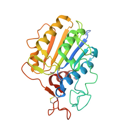
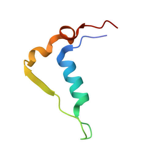
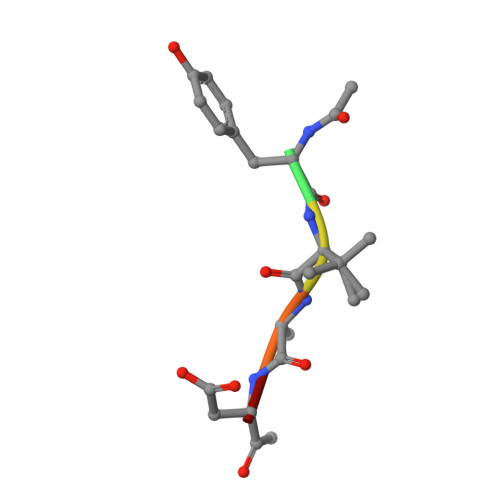
Structural analyses ofArabidopsis thalianalegumain gamma reveal differential recognition and processing of proteolysis and ligation substrates.

Zauner, F.B.Elsasser, B.Dall, E.Cabrele, C.Brandstetter, H.

(2018) J Biological Chem 293: 8934-8946

  • DOI: https://doi.org/10.1074/jbc.M117.817031
  • Primary Citation Related Structures: 
    5OBT

  • PubMed Abstract: 

    Legumain is a dual-function protease-peptide ligase whose activities are of great interest to researchers studying plant physiology and to biotechnological applications. However, the molecular mechanisms determining the specificities for proteolysis and ligation are unclear because structural information on the substrate recognition by a fully activated plant legumain is unavailable. Here, we present the X-ray structure of Arabidopsis thaliana legumain isoform γ (AtLEGγ) in complex with the covalent peptidic Ac-YVAD chloromethyl ketone (CMK) inhibitor targeting the catalytic cysteine. Mapping of the specificity pockets preceding the substrate-cleavage site explained the known substrate preference. The comparison of inhibited and free AtLEGγ structures disclosed a substrate-induced disorder-order transition with synergistic rearrangements in the substrate-recognition sites. Docking and in vitro studies with an AtLEGγ ligase substrate, sunflower trypsin inhibitor (SFTI), revealed a canonical, protease substrate-like binding to the active site-binding pockets preceding and following the cleavage site. We found the interaction of the second residue after the scissile bond, P2'-S2', to be critical for deciding on proteolysis versus cyclization. cis-trans -Isomerization of the cyclic peptide product triggered its release from the AtLEGγ active site and prevented inadvertent cleavage. The presented integrative mechanisms of proteolysis and ligation (transpeptidation) explain the interdependence of legumain and its preferred substrates and provide a rational framework for engineering optimized proteases, ligases, and substrates.


  • Organizational Affiliation
    • From the Department of Biosciences, University of Salzburg, Salzburg 5020, Austria.

Macromolecule Content 

  • Total Structure Weight: 66.56 kDa 
  • Atom Count: 5,177 
  • Modeled Residue Count: 570 
  • Deposited Residue Count: 604 
  • Unique protein chains: 3

Macromolecules

Find similar proteins by:|  3D Structure
Entity ID: 1
MoleculeChains  Sequence LengthOrganismDetailsImage
Vacuolar-processing enzyme gamma-isozymeA,
D [auth B]
245Arabidopsis thalianaMutation(s): 0 
Gene Names: At4g32940F26P21.60
EC: 3.4.22.34
UniProt
Find proteins for Q39119 (Arabidopsis thaliana)
Explore Q39119 
Go to UniProtKB:  Q39119
Entity Groups
Sequence Clusters30% Identity50% Identity70% Identity90% Identity95% Identity100% Identity
UniProt GroupQ39119
Sequence Annotations
Expand
Reference Sequence
Find similar proteins by:|  3D Structure
Entity ID: 2
MoleculeChains  Sequence LengthOrganismDetailsImage
Vacuolar-processing enzyme gamma-isozymeB [auth E],
E [auth F]
51Arabidopsis thalianaMutation(s): 0 
Gene Names: At4g32940F26P21.60
EC: 3.4.22.34
UniProt
Find proteins for Q39119 (Arabidopsis thaliana)
Explore Q39119 
Go to UniProtKB:  Q39119
Entity Groups
Sequence Clusters30% Identity50% Identity70% Identity90% Identity95% Identity100% Identity
UniProt GroupQ39119
Sequence Annotations
Expand
Reference Sequence
Find similar proteins by:  Sequence   |   3D Structure  
Entity ID: 3
MoleculeChains  Sequence LengthOrganismDetailsImage
Ac-YVAD-CMKC,
F [auth D]
6synthetic constructMutation(s): 0 

Small Molecules

Modified Residues  1 Unique
IDChains TypeFormula2D DiagramParent
SNN
Query on SNN
A,
D [auth B]
L-PEPTIDE LINKINGC4 H6 N2 O2ASN

Biologically Interesting Molecules (External Reference) 

1 Unique

Experimental Data & Validation

Experimental Data

  • Method: X-RAY DIFFRACTION
  • Resolution: 1.50 Å
  • R-Value Free:  0.182 (Depositor), 0.182 (DCC) 
  • R-Value Work:  0.152 (Depositor), 0.153 (DCC) 
  • R-Value Observed: 0.153 (Depositor) 
Space Group: P 1 21 1
Unit Cell:
Length ( Å )Angle ( ˚ )
a = 41.74α = 90
b = 78.07β = 92.98
c = 77.719γ = 90
Software Package:
Software NamePurpose
PHENIXrefinement
iMOSFLMdata reduction
Aimlessdata scaling
PHASERphasing

Structure Validation

View Full Validation Report


Entry History 

& Funding Information

Deposition Data


Funding OrganizationLocationGrant Number
Austrian Science FundAustriaW_01213

Revision History  (Full details and data files)

  • Version 1.0: 2018-04-11
    Type: Initial release
  • Version 1.1: 2018-04-18
    Changes: Data collection, Database references
  • Version 1.2: 2018-06-13
    Changes: Data collection, Structure summary
  • Version 1.3: 2018-06-20
    Changes: Data collection, Database references
  • Version 2.0: 2023-11-15
    Changes: Atomic model, Data collection, Database references, Derived calculations
  • Version 2.1: 2024-01-17
    Changes: Refinement description
  • Version 2.2: 2024-11-06
    Changes: Structure summary