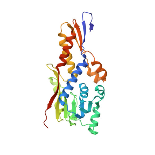
5O4N | pdb_00005o4n

Apo HcgC from Methanococcus maripaludis soaked with SAH and pyridinol


Experimental Data Snapshot

  • Method: X-RAY DIFFRACTION
  • Resolution: 2.05 Å
  • R-Value Free: 
    0.236 (Depositor), 0.230 (DCC) 
  • R-Value Work: 
    0.197 (Depositor), 0.200 (DCC) 
  • R-Value Observed: 
    0.199 (Depositor) 

Starting Model: experimental
View more details

wwPDB Validation   3D Report Full Report


Ligand Structure Quality Assessment 

Created with Raphaël 2.3.0Worse 01 BetterLigand structure goodness of fit to experimental dataBest fitted SAHClick on this verticalbar to view detailsBest fitted 9KHClick on this verticalbar to view details

This is version 1.3 of the entry. See complete history


Literature

A Water-Bridged H-Bonding Network Contributes to the Catalysis of the SAM-Dependent C-Methyltransferase HcgC.

Bai, L.Wagner, T.Xu, T.Hu, X.Ermler, U.Shima, S.

(2017) Angew Chem Int Ed Engl 56: 10806-10809

  • DOI: https://doi.org/10.1002/anie.201705605
  • Primary Citation of Related Structures:  
    5O4H, 5O4J, 5O4M, 5O4N

  • PubMed Abstract: 

    [Fe]-hydrogenase hosts an iron-guanylylpyridinol (FeGP) cofactor. The FeGP cofactor contains a pyridinol ring substituted with GMP, two methyl groups, and an acylmethyl group. HcgC, an enzyme involved in FeGP biosynthesis, catalyzes methyl transfer from S-adenosylmethionine (SAM) to C3 of 6-carboxymethyl-5-methyl-4-hydroxy-2-pyridinol (2). We report on the ternary structure of HcgC/S-adenosylhomocysteine (SAH, the demethylated product of SAM) and 2 at 1.7 Å resolution. The proximity of C3 of substrate 2 and the S atom of SAH indicates a catalytically productive geometry. The hydroxy and carboxy groups of substrate 2 are hydrogen-bonded with I115 and T179, as well as through a series of water molecules linked with polar and a few protonatable groups. These interactions stabilize the deprotonated state of the hydroxy groups and a keto form of substrate 2, through which the nucleophilicity of C3 is increased by resonance effects. Complemented by mutational analysis, a structure-based catalytic mechanism was proposed.


  • Organizational Affiliation

    Max-Planck-Institut für terrestrische Mikrobiologie, Karl-von-Frisch-Straße 10, 35043, Marburg, Germany.


Macromolecules
Find similar proteins by:  (by identity cutoff)  |  3D Structure
Entity ID: 1
MoleculeChains Sequence LengthOrganismDetailsImage
HcgC
A, B, C, D
274Methanococcus maripaludis S2Mutation(s): 0 
Gene Names: MMP1498
UniProt
Find proteins for Q6LX54 (Methanococcus maripaludis (strain DSM 14266 / JCM 13030 / NBRC 101832 / S2 / LL))
Explore Q6LX54 
Go to UniProtKB:  Q6LX54
Entity Groups  
Sequence Clusters30% Identity50% Identity70% Identity90% Identity95% Identity100% Identity
UniProt GroupQ6LX54
Sequence Annotations
Expand
  • Reference Sequence
Small Molecules
Ligands 4 Unique
IDChains Name / Formula / InChI Key2D Diagram3D Interactions
SAH
Query on SAH

Download Ideal Coordinates CCD File 
E [auth A],
I [auth B],
M [auth C],
S [auth D]
S-ADENOSYL-L-HOMOCYSTEINE
C14 H20 N6 O5 S
ZJUKTBDSGOFHSH-WFMPWKQPSA-N
9KH
Query on 9KH

Download Ideal Coordinates CCD File 
F [auth A],
J [auth B],
N [auth C],
T [auth D]
6-carboxy methyl-4-hydroxy-2-pyridinol
C8 H9 N O4
MGGXBHMHJPCYSF-UHFFFAOYSA-N
MPD
Query on MPD

Download Ideal Coordinates CCD File 
G [auth A],
H [auth A],
K [auth B],
O [auth C],
U [auth D]
(4S)-2-METHYL-2,4-PENTANEDIOL
C6 H14 O2
SVTBMSDMJJWYQN-YFKPBYRVSA-N
DMS
Query on DMS

Download Ideal Coordinates CCD File 
L [auth B],
P [auth C],
Q [auth C],
R [auth C]
DIMETHYL SULFOXIDE
C2 H6 O S
IAZDPXIOMUYVGZ-UHFFFAOYSA-N
Experimental Data & Validation

Experimental Data

  • Method: X-RAY DIFFRACTION
  • Resolution: 2.05 Å
  • R-Value Free:  0.236 (Depositor), 0.230 (DCC) 
  • R-Value Work:  0.197 (Depositor), 0.200 (DCC) 
  • R-Value Observed: 0.199 (Depositor) 
Space Group: P 1 21 1
Unit Cell:
Length ( Å )Angle ( ˚ )
a = 72.556α = 90
b = 80.997β = 108.24
c = 97.808γ = 90
Software Package:
Software NamePurpose
BUSTERrefinement
XDSdata reduction
SCALAdata scaling
MOLREPphasing

Structure Validation

View Full Validation Report


Ligand Structure Quality Assessment 

Created with Raphaël 2.3.0Worse 01 BetterLigand structure goodness of fit to experimental dataBest fitted SAHClick on this verticalbar to view detailsBest fitted 9KHClick on this verticalbar to view details

Entry History 

Deposition Data

Revision History  (Full details and data files)

  • Version 1.0: 2017-07-19
    Type: Initial release
  • Version 1.1: 2017-08-09
    Changes: Database references
  • Version 1.2: 2017-09-06
    Changes: Database references
  • Version 1.3: 2024-01-17
    Changes: Data collection, Database references, Refinement description